ABSTRACT
Background: Dermatophytoses are one of the commonest skin affections encountered in tropical regions and especially in India. Most common of them being the tinea corporis i.e., the fungal infection of the body other than scalp, fingers, nails, foot and groin regions. The condition is generally not life threatening but affects the physical, mental and social quality of affected individual. The conventional medical treatment considers it as superficial disorder generally and treats in the same direction with topical agents in most of the cases. Most of the times causing either re -appearance of the lesions at different site or suppression. Homoeopathy offers gentle and permanent way to treat this condition. It considers the patient’s condition holistically, and treating the same. The miasmatic concept of disease is one of the tenets of homoeopathic philosophy which helps in understanding the disease development and in its cure, this concept demands to be further researched.
INTRODUCTION
TINEA CORPORIS
Dermatophytes are fungi that invade and multiply within keratinized tissues (skin, hair, and nails) causing infection. Based upon their genera, dermatophytes can be classified into three groups: Trichophyton (which causes infections on skin, hair, and nails), epidermophyton (which causes infections on skin and nails), and Microsporum (which causes infections on skin and hair).Tinea corporis, also known as ‘ringworm,’ is a superficial dermatophyte infection of the skin, other than on the hands (tinea manuum), feet (tinea pedis), scalp (tinea capitis), bearded areas (tinea barbae), face (tinea faciei), groin (tinea cruris), and nails (onychomycosis or tinea unguium).Dermatophytosis has a significant impact on the patients’ quality of life by affecting psychological, economic, and social aspects, and are associated with anxiety, depression, and low self-esteem, mainly due to discomfort regarding pruritus and cosmetic issues.
HISTORY
In the past, a number of diseases used to be lumped under a common expression. The terms like lichen, lepra, lupus, herpes, psora are few examples. Ringworm was no exception. Various diseases, particularly of annular configuration, were put together under some common headings. Guy de Chauliac (AD 1300–1368), a French physician, used the word Tinea as a generic term for many such ailments. Willan replaced it by Porrigo scutulata. Alibert classified tinea and his Tigne granulée was again a doubtful entity. Other authors designated it in various forms like Herpes squamosus (Cazenev), Tineatondante (Mahon), Phyto-alopecia (Melmsten), Tinea circinata (Anderson), Trichinosis furfuracea (Devergie) to name a few. The early discussions were centered around the scalp ringworms and the most used designation was Favus. Other terms used were Porrigo scutulata, Trichophytica capitis, Tinea ficosa, Achores even Scabies capitis simplex (Plenck). Even David Gruby, the pioneer of dermatophytological research, named it Porrigo decalvans – a term already used by Bateman for alopecia areata – an altogether different disease. In 1835, Rayer in his famous ‘A theoretical and practical treatise on the diseases of the skin’ described ringworm under the banner Impetigo annulata. Schamberg in 1896 also lumped ringworm with some other diseases under the title Impetigo contagiosa annulata et serpiginosa. The confusion also hovered around the etiology. It was believed that a common organism was responsible for different varieties of ringworms. This trend went on till 1894, when Sabouraud published his notable findings in Les Trichophyties Humaines, avec Atlas establishing the plurality of the etiological agent of ringworm. In 1910, Sabouraud published his magnum opus Les Tiegnes and a new era begun.
More than a century has elapsed since then and we still are facing the riddle as to the peculiar behaviour of the ringworms every moment at present. Much work is yet to be done. There is a long way to go before we sleep.
EPIDEMIOLOGY
Tinea corporis is the most common dermatophytosis.While tinea corporis occurs worldwide, it is most commonly observed in tropical regions.The lifetime risk of acquiring tinea corporis is estimated to be 10–20%.The current reported prevalence in India falls in a very wide range (6.09%6– 61.5%7). A prevalence of 6.09% to 27.6% has been reported in studies from south India,6.8 while a high prevalence of 61.5% has been recorded in north India. there appears to be a steady increase in the incidence of chronic, relapsing and recurrent dermatophytosis; it is not uncommon to encounter disease durations running into months or years.
Age and sex
Tinea corporis occurs most frequently in post-pubertal children and young adults. Rare cases have been reported in the newborn period. There is no sex predominance. Chronic dermatophytosis (disease for more than 6 months with or without recurrence despite being treated) is more common in the late middle age group, around fifth decade, which is attributed to waning immunity, comorbidities like diabetes and other risk factors such as positive family history, use of topical steroid-antifungal creams, immunosuppressants intake etc.
Familial cases
Transmission among household family members is by far the most common route; children often become infected by spores shed by an infected household family member. Chronicity and recurrent infection have probably contributed to the rise in the incidence of familial infection. Connubial dermatophytosis is common. A high prevalence occurs in people living in overcrowded homes, slums, hostel rooms and dormitories.
Urban versus rural areas and literacy
People from both urban and rural areas are at increased risk of dermatophytic infections. Studies from the first half of the past decade reported a rural predominance, possibly due to the high frequency of outdoor work including agriculture predisposing to increased perspiration. However, studies in the last 5 years have shown greater proportions of patients from urban areas (around 80% of patients). Education and literacy have not been commented upon much in reports concerning the increased incidence of dermatophytosis.
Socioeconomic status
A higher proportion of patients with dermatophyte infections are still from lower socioeconomic groups, with studies reporting an incidence of 61–67%. This is followed by lower-middle and medium socioeconomic strata.9.19,26 Poor standards of living, lack of hygiene, overcrowding and poor nutrition in the lower socioeconomic groups promote the growth of dermatophytes, increasing the risks of infection, chronicity and recurrence.
Occupation
People engaged in outdoor activities in hot and humid environments are at a greater risk of infection since this provides a favourable environment for dermatophytes. Recent studies too have reported that manual labourers are most commonly affected. Farmers are at an additional risk due to increased exposure to fungal pathogens from the environment and frequent contact with soil and animals. The hot environment of the kitchen with increased sweating favors the growth of dermatophytes, making homemakers susceptible. An increased frequency of dermatophytosis has also been reported in students recently.
Topical steroid abuse
Topical steroid misuse may be the most important cause of the current outbreak of chronic and recalcitrant dermatophytosis. A strong temporal association has been observed between the increasing availability and irrational use of the combination creams (antifungal-steroid or antifungal-antibiotic) with the sudden increase in chronic, recurrent and refractory cases in the past 4–5 years.
ETIOLOGY
Tinea corporis is most often caused by Trichophyton rubrum, T. tonsurans, and Microsporum canis. T. rubrum is by far the most common cause of dermatophytosis worldwide. Tinea corporis secondary to tinea capitis is often caused by T. tonsurans.16 On the other hand, tinea corporis resulting from close contact with dogs or cats is often caused by M. canis.8,17–19 Other causative organisms include T. interdigitale (previously known as T. mentagrophytes), T. verrucosum, T. violaceum, T. concentricum, Epidermophyton floccosum, M. audouinii, and M. gypseum. In recent years, T. interdigitale has replaced T. rubrum as the most common cause of tinea corporis in Southeast Asia.
PATHOGENESIS
Mannans in the cell walls of some dermatophytes, such as T. rubrum, have immune-inhibitory properties. This allows the fungus to stay on the skin without being sloughed off prior to invasion of the skin. The causative fungus can produce proteases (enzymes that digest keratin), serine-subtilisins (enzymes that digest protein by initiating the nucleophilic attack on the peptide bond through a serine residue at the active site), and keratinases (enzymes that penetrate keratinized tissue), which allow the fungus to invade the horny layer of the skin and spread outward. Infection is usually cutaneous and confined to the outer, non-living, cornified layers of the skin. The fungus is unable to penetrate the deeper tissues in healthy immunocompetent hosts because of host defense mechanisms, such as activation of serum inhibitory factor, polymorphonuclear leukocytes, and complements. Scaling of the active border results from increased epidermal cell proliferation in response to the fungal infection.11,17,18
CLINICAL MANIFESTATIONS
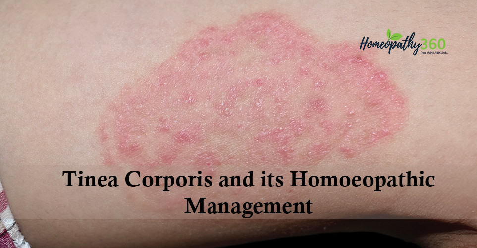
The incubation period is 1–3 weeks. Tinea corporis typically presents as a well-demarcated, sharply circumscribed, oval or circular, mildly erythematous, scaly patch or plaque with a raised leading edge. The lesion starts off as a flat scaly spot that spreads centrifugally and clears centrally to form a characteristic annular lesion giving rise to the term ‘ringworm. The central area becomes hypopigmented or brown and less scaly as the active border progresses outward. The border is usually annular and irregular. Occasionally, the border can be papular, vesicular, or pustular. Lesions may assume other shapes such as circinate and arcuate. Mild pruritus is common. In general, lesions caused by anthropophilic species (e.g. T. rubrum, T. tonsurans, T. interdigitale, T. schoenleinii, T. soundanense, T. violaceum, M. audouinii, and E. floccosum) are often less inflammatory/ erythematous than those caused by zoophilic species or geophilic species. The lesions tend to be asymmetrically distributed. When multiple lesions are present, they may coalesce into polycyclic patterns. In adults, tinea corporis most commonly occurs on exposed skin. In children and adolescents, the site of predilection is the trunk. In tinea gladiatorum, the lesion presents as well-demarcated, erythematous, annular, scaling plaques on areas of skin-to-skin contact, such as the head, neck, and arms. Tinea gladiatorum is most often caused by T. tonsurans. The condition is most common among those who engage in contact sports such as wrestling and judo. Tinea incognito refers to a cutaneous fungal infection that has lost its classical morphological features because of the use of calcineurin inhibitors or corticosteroids. The clinical manifestations of tinea incognito are highly variable. Generally, compared with the lesion of tinea corporis, the lesion seen in tinea incognito is less erythematous and scaly, with a less defined border and is typically more widespread. Pruritus is usually mild or absent. The rash can be eczema-like, rosacea-like, or discoid lupus erythematosus-like, especially on the face, and eczema-like or impetigo-like on the trunk and limbs.
Many clinical variants of tinea corporis exist. Tinea imbricata, caused mainly by a strictly anthropophilic dermatophyte, T. concentricum, typically presents as multiple, scaly, annular, concentric, erythematous rings that can extend to form polycyclic plaques. With time, multiple overlapping lesions develop and the plaques become lamellar with abundant thick scales adhering to the interior of the plaque, giving rise to the appearance of overlapping roof tiles or lace, fish scales. The trunk is the site of predilection. Tinea imbricata has a high tendency to generalize and large areas of the body may be affected. Pruritus is common. Tinea imbricata is endemic in Central and South America, Southwest Pacific, and Southeast Asia.
DIAGNOSIS
The diagnosis of tinea corporis is most often clinical, especially if the lesion is typical.19 A well-demarcated, sharply circumscribed, erythematous, annular, scaly plaque with a raised leading edge, and scaling and central clearing on the body is characteristic. At times, the diagnosis can be difficult due to the prior use of medications, such as calcineurin inhibitors or corticosteroids. Dermoscopy is a useful and non-invasive diagnostic tool. Dermoscopic findings in cases of tinea corporis include diffuse erythema, dotted vessels with peripheral to patchy distribution, white scales with peripheral distribution, ‘moth-eaten’ scale, peeling in an outward direction, brown spots surrounded by a white-yellow halo, follicular micro-pustules, wavy hair, and broken hair. These changes may be seen despite the use of topical corticosteroids or calcineurin inhibitors. Reflectance confocal microscopy is another useful diagnostic tool. With reflectance confocal microscopy, branching fungal hyphae can be detected over an erythematous annular scaly patch in individuals with tinea corporis. Wood lamp examination of the affected area is not useful as the lesion of tinea corporis usually does not fluoresce with a Wood lamp.20-23
If necessary, the diagnosis can be confirmed by microscopic examination of potassium hydroxide (KOH) wet-mount preparations of skin scrapings from the active border of the lesion. The skin scrapings should be transported in a presterilized black chart paper so as to keep the specimen dry and prevent overgrowth of bacteria that may be contaminants. To, perform the test, a drop of 10–20% KOH is added to the scrapings on a microscopic slide. The specimen is gently heated to accelerate the destruction of the squamous cells. The KOH dissolves the epithelial tissue, leaving behind easily visualized septate hyphae with or without arthro-conidiospores. The addition of dimethyl sulfoxide to KOH may permit more rapid examination without heating.
Fungal culture is the gold standard to diagnose dermatophytosis, especially if the diagnosis is in doubt and results of other tests are inconclusive, or the infection is widespread, severe, or unresponsive to treatment. Fungal culture can help to differentiate fungal species. However, fungal culture is expensive and it usually takes 7–14 days for results.12,18,24,25
DIFFERENTIAL DIAGNOSIS
Diseases that present with annular lesions may mimic tinea corporis. The differential diagnosis is broad and includes wide variety of lesions.
Pityriasis rosea (non-itchy herald patch, generalized, bilateral, symmetrical eruption 4–14 days later, characteristic ‘Christmas tree’ appearance on the back and a V-shaped pattern on the upper chest);
Tinea versicolor (multiple, well-demarcated, finely scaly, brownish macules/patches in fair-skinned individuals and hypopigmented macules/patches in dark-skinned individuals, minimal or absent erythema, absent collarette of scales in individual lesions, typically asymptomatic);
Nummular eczema (well-demarcated, pruritic, coin-shaped, symmetrical, eczematous, scaly lesions, involvement of the extremities rather than the trunk, serous exudate in acute lesions, no central clearing, rapid response to topical steroids);
Plaque psoriasis (well-demarcated, sharply circumscribed, annular, erythematous, round or oval, pruritic plaques with loosely adherent silvery-white micaceous scales, positive Auspitz sign, Koebner phenomenon, nail pitting, arthritis, uveitis, geographic tongue, positive family history);
Atopic Dermatitis (flexural involvement in older children and adolescents, highly pruritic, excoriations, lichenification in chronic lesions, chronically relapsing);
Contact Dermatitis (well-demarcated, erythematous lesion localized to the area of contact, immediate skin reaction with burning, stinging, or discomfort if caused by an irritant, delayed response associated with pruritus caused by an allergen);
Seborrheic Dermatitis (salmon-colored or erythematous, sharply demarcated patches with yellow-white, greasy scales);
Localized Granuloma Annulare (asymptomatic, firm, erythematous, violaceous, flesh-colored or brown, non-scaly plaques with central involution, annular configuration, usually involve the extensor surfaces of distal extremities);
Fixed drug eruption;
Subacute Cutaneous Lupus Erythematosus (annular, erythematous, scaly plaques often in sun-exposed areas);
Discoid Lupus Erythematosus (well-demarcated, erythematous, hyperkeratotic, indurated, coin-shaped plaques covered bypartially adherent, scales in sun-exposed areas);
Urticaria (pruritic, erythematous, and edematous wheals of the superficial layers of the skin, individual lesions wax and wane rapidly);
Lichen Planus (characterized by 6 Ps: planar (flat-topped), purple (violaceous), polygonal, pruritic, papules/ plaques, lesions may be covered with white, lacy, reticular lines [Wickham striae]);
Secondary Syphilis (asymptomatic, diffuse, symmetrical, round-to-oval, pink-to-reddish-brown mono-morphos macules or patches on the trunk and extremities including the palms and soles, absence of herald patch, ‘moth-eaten’ alopecia, lymphadenopathy, history of venereal exposure, and/or chancre).
HOMOEOPATHIC VIEWPOINT IN TINEA CORPORIS
The trichophyton is not the disease itself, but its organic scavenger. Cure the internal disease, and this scavenger dies. – J. C. BURNETT
Homoeopathic philosophy considers man as whole is diseased not any single part or organ is diseased. It considers that the life principle of the living organism must be deranged first by inimical agent, resulting in development of varying pathologies in man. As said by Kent, that man is the will and understanding and the house which he lives in is his body, man is prior to the organ and it is man who is sick prior to the body.
Tinea corporis is considered as superficial disease of outer covering of human being i.e., skin and treated in the same direction but homoeopathic comprehension of this disease is different, J H Allen says about skin diseases that skin is the mirror or the reflector of the internal stress, the internal dynamis, the internal workings of this human machine. It has in the skin, its reflectors, its kaleidoscope, its kinetoscopic views of its internal movements, and its multiple shadings of disease, its lights and its shadows that go to make up a picture, thrown upon that human canvas, the skin, showing much of perverted life action in the organism. All skin eruptions are either secondary or tertiary expressions of miasmatic action. Pathologically speaking, we look upon the outer man for signs, for markings or pencillings that tell of the kind of life within the organism itself. Sometimes these pencillings are like shadowgraphs, showing only faint tracings of the presence of a latent miasm, and again they may be well defined and well developed even to physiological changes of form, color and proportions.
When se look upon these lesions of the skin as local states or changes in itself, we simply ignore that co-operative principles that rules throughout the organism as a whole, and we attribute that power to a part and not to that which governs the whole. Therefore, our therapeutic efforts are themselves misdirected and instead of directing the perverted life forces aright, we misguide them, bringing about nothing but Babylon or confusion.
Dr. Hahnemann has of view, about this concept of considering skin diseases as local, is highlighted in aphorisms of Organon of medicine as – Among the one-sided disease an important place is occupied by the so-called local maladies, by which term is signified those changes and ailments that appear on the external parts of the body. Till now the idea prevalent in the schools was that these parts were alone morbidly affected, and that the rest of the body did not participate in the disease – a theoretical, absurd doctrine, which has led to the most disastrous medical treatment. (§185).
He further mentions about different aspects of these in different aphorism as follows6:
Aphorism § 188: These affections were considered to be merely topical, and were therefore called local diseases, as if they were maladies exclusively limited to those parts wherein the organism took little or no part, or affections of these particular visible parts of which the rest of the living organism, so to speak, knew nothing.
Aphorism § 189: And yet very little reflection will suffice to convince us that no external malady (not occasioned by some important injury from without) can arise, persist or even grow worse without some internal cause, without the co-operation of the whole organism, which must consequently be in a diseased state. It could not make its appearance at all without the consent of the whole of the rest of the health, and without the participation of the rest of the living whole (of the vital force that pervades all the other sensitive and irritable parts of the organism); indeed, it is impossible to conceive its production without the instrumentality of the whole (deranged) life; so intimately are all parts of the organism connected together to form an indivisible whole in sensation and functions. No eruption on the lips, no whitlow can occur without previous and simultaneous internal ill-health.
Aphorism § 190: All true medical treatment of a disease on the external parts of the body that has occurred from little or no injury from without must, therefore, be directed against the whole, must effect the annihilation and cure of the general malady by means of internal remedies, if it is wished that the treatment should be judicious, sure, efficacious and radical.
Aphorism § 194: It is not useful, either in acute local diseases of recent origin or in local affections that have already existed a long time, to rub in or apply externally to the spot an external remedy, even though it be the specific and, when used internally, salutary by reason of its homoeopathicity, even although it should be at the same time administered internally; for the acute topical affections (e.g., inflammations of the individual parts, erysipelas, etc.), which have not been caused by external injury of proportionate violence, but by dynamic or internal causes, yield most surely to internal remedies homoeopathically adapted to the perceptible state of the health present in the exterior and interior, selected from the general store of proved medicines,1 and generally without any other aid; but if these diseases do not yield to them completely, and if there still remain in the affected spot and in the whole state, notwithstanding good regimen, a relic of disease which the vital force is not competent to restore to the normal state, then the acute disease was (as not infrequently happens) a product of psora which had hitherto remained latent in the interior, but has now burst forth and is on the point of developing into a palpable chronic disease.
Aphorism § 198: The mere topical employment of medicines, that are powerful for cure when given internally, to the local symptoms of chronic miasmatic diseases is for the same reason quite inadmissible; for if the local affection of the chronic disease be only removed locally and in a one-sided manner, the internal treatment indispensable for the complete restoration of the health remains in dubious obscurity; the chief symptom (the local affection) is gone, and there remain only the other, less distinguishable symptoms, which are less constant and less persistent than the local affection, and frequently not sufficiently peculiar and too slightly characteristic to display after that, a picture of the disease in clear and peculiar outlines.
Aphorism § 201: It is evident that man’s vital force, when encumbered with a chronic disease which it is unable to overcome by its own powers, adopts the plan of developing a local malady on some external part, solely for this object, that by making and keeping in a diseased state this part which is not indispensable to human life, it may thereby silence the internal disease, which otherwise threatens to destroy the vital organs (and to deprive the patient of life), and that it may thereby, so to speak, transfer the internal disease to the vicarious local affection and, as it were, draw it thither. The presence of the local affection thus silences, for a time, the internal disease, though without being able either to cure it or to diminish it materially.1 The local affection, however, is never anything else than a part of the general disease, but a part of it increased all in one direction by the organic vital force, and transferred to a less dangerous (external) part of the body, in order to allay the internal ailment. But (as has been said) by this local symptom that silences the internal disease, so far from anything being gained by the vital force towards diminishing or curing the whole malady, the internal disease, on the contrary, continues, in spite of it, gradually to increase and Nature is constrained to enlarge and aggravate the local symptom always more and more, in order that it may still suffice as a substitute for the increased internal disease and may still keep it under. Old ulcers on the legs get worse as long as the internal psora is uncured, the chancre enlarges as long as the internal syphilis remains uncured, just as the general internal disease continues to increase as times goes on.
Aphorism §204: If we deduct all chronic affections, ailments and diseases that depend on a persistent unhealthy mode of living, (§ 77) as also those innumerable medicinal maladies (v. § 74) caused by the irrational, persistent, harassing and pernicious treatment of diseases often only of trivial character by physicians of the old school, all the remainder, without exception, result from the development of these three chronic miasms, internal syphilis, internal sycosis, but chiefly and in infinitely greater proportion, internal psora, each of which was already in possession of the whole organism, and had penetrated it in all directions before the appearance of the primary, vicarious local symptom of each of them (in the case of psora the scabious eruption, in syphilis the chancre or the bubo, and in sycosis the condylomata) that prevented their outburst; and these chronic miasmatic diseases, if deprived of their local symptom, are inevitably destined by mighty Nature sooner or later to become developed and to burst forth, and thereby propagate all the nameless misery, the incredible number of chronic diseases which have plagued mankind for hundreds and thousands of years, none of which would so frequently have come into existence had physicians striven in a rational manner to cure radically and to extinguish in the organism these three miasms by the internal homoeopathic medicines suited for each of them, without employing topical remedies for their external symptoms.
Homoeopathy has insightful and deep understanding towards the treatment of skin diseases. Homeopathy strongly believes in understanding the holistic causative factors while handling skin diseases, whereby the study of skin, mind, constitution, miasmatic background, genetic influences, domestic and social environment are put together to determine the line of treatment.
Ringworm being a true chronic disease, miasmatic factors are responsible for it and selected medicine must have similar miasmatic semblance. At the same time, it is equally necessary to give importance on accessory circumstances whether these things have tended to increase the malady or not and if present it necessary to remove or modify them.
HOMOEOPATHIC THERAPEUTICS FOR TINEA CORPORIS30,31
Arsenicum album: It has sensitiveness to disorderliness or untidiness. Anxiety and fear of death. Restless disposition. Extremely chilly individual, hugs the fire in the winter, wants to be well wrapped up. Thirst is characteristic, with a desire for frequent sips of water. Prefers warm or hot drinks. Aggravated from Cold food, cold drink, cold air, wet weather. Skin: dry, scaly, bran like, dirty, white skin; herpetic eruption with itching and burning; Burning sensation in lesions. Burning pain relieved by heat. Tettery spots, covered with phlyctenae and furfur, with burning nocturnal pains.
Bacillinum: Dr. Burnett has shown that ringworm of the scalp and pityriasis versicolor on the body are indications of tubercular diathesis, and they respond to this remedy. Also, they are leading indications for it when present in combination with other affections. Ringworm. Worse, night and early morning; cold air.
CHRYSAROBINUM: Acts as a powerful irritant of the skin and used successfully in skin diseases especially in ringworm, psoriasis, herpes tonsurans acne rosacea. Violent itching, thighs, legs and ears. Dry, scaly eruption, especially around eyes and ears, scabs with pus underneath.
Dulcamara: Chilly patient. Catarrhal skin affections brought on or aggravated by exposure to cold, damp, rainy weather, or sudden changes in hot weather. Precipitating causes are suppressed sweats, exposure to cold, working while standing in cold water. Humid eruptions on cheek. Thick, brown, herpetic crusts on the face, forehead, temples and chin with reddish borders, bleeding when scratched. Agg. in cold wet weather. Better by external warmth. Tetters of different kinds, such as humid, scaly, pale tetters, oozing after having been scratched; reddish tetters, with red areola, bleeding after having been scratched; tetters with red edges, painfully sensitive to the touch, and to cold water; small, round, yellowish-brown tetters, bleeding after having been scratched; dry, furfuraceous tetters.
Graphitis: It is best suited to fatty, flabby, chilly and costive person. Extreme hesitation, unable to make up her mind about anything. Chilly. Skin inclined to crack. Fissures deep, bleeding or oozing out a sticky fluid. Eruptions upon the ears, between fingers and toes and on various parts of body, from which oozes a watery, transparent, sticky fluid. Warmth of bed aggravates itching. Tetters, and other humid or scabby eruptions, sometimes with secretion of corrosive serum, or with itching in the evening, and at night.
Natrum muriaticum: It covers all three Miasms. Suited to anaemic, chlorotic and emaciated person, emaciation most marked about the neck, which is very thin and shrunken. Herpes about anus and on borders of hair at nape of neck. Eczema: raw, red, inflamed, especially in edges of hair; < from eating too much salt, at sea shore, or from ocean voyage.
Natrium sulphuricum: Chilly patient. Diseases induced by damp weather or living in damp houses; patient feels every change of weather, especially from dry to wet. Every spring skin affections reappear. Itching while undressing. Violent itching in genital organs, of scrotum, of perineum, of mons veneris with burning after scratching. Violent itching of toes, and between toes, especially on taking off shoes and stocking at night.
Petroleum: Herpes: of genital organs extending to perineum and thighs; itching, redness; skin cracked, rough, bleeding; dry or moist. The skin symptoms are worse in winter, better in summer. All eruption itches violently. Cannot rest until he scratches the skin off, when the part becomes moist, bloody, raw and inflamed.
Sepia: Suited to pot-bellied mothers, yellow saddle across nose, irritable, women. Skin diseases developing on brunettes who suffer from chronic uterine troubles. Herpes circinatus in isolated spots on upper part of body. Itching of skin; of various parts; of external genitalia; is not > by scratching, and is apt to change to burning. Herpetic eruption on lips, about mouth and nose. Ringworm-like eruption every spring. Moist, scabious herpes, with itching and burning sensation.
Sulphur: Persons who are lean, stooped-shouldered and walk and sit stooped. Orifice of body very red, lips bright red. Skin: itching, voluptuous; scratching >; “feels good to scratch (voluptuous itching)”; Scratching causes burning; < from heat of bed, washing, at night.
Tellurium metallicum: Herpes circinatus in intersecting rings over whole body. Body thickly covered with elevated rings of herpes circinatus. Itching of hands and feet. Herpetic spots; ringworm. Ring-shape lesions, offensive odours from affected parts. Barber’s itch. Ringworm worse on lower extremities.
Thuja: Dry skin, with brown spots. Herpetic eruptions. Tearing pains in glands. Glandular enlargement. Nails crippled; brittle and soft. Eruptions only on covered parts; worse after scratching. Very sensitive to touch. Coldness of one side. Brown spots on hands and arms. Worse, at night, from heat of bed; at 3 am and 3 pm; from cold, damp air. The majority of cutaneous symptoms are > by touch.
Tuberculinum: It is indicated in light complexion person with blue eyes, suffering from a tubercular diathesis. Loses flesh while eating well. Itching intense, < at night when undressing, from bathing; immense quantities of white bran-like scales; oozing behind the ears, in the hair, in folds of skin with rawness and soreness; fiery red skin. Itching all over the body in the evening in bed; changing place after rubbing.