Authored by:
Dr Vishnu T M
Assistant Professor, Dept of Anatomy
Yenepoya Homoeopathic Medical College and HospitalMangalore.
Kidney stones (also called renal calculi, nephrolithiasis or urolithiasis) are hard deposits made of minerals and salts that form inside your kidneys. Diet, excess body weight, some medical conditions, and certain supplements and medications are among the many causes of kidney stones. Kidney stones can affect any part of your urinary tract from your kidneys to your bladder. Often, stones form when the urine becomes concentrated, allowing minerals to crystallize and stick together. Passing kidney stones can be quite painful, but the stones usually cause no permanent damage if they’re recognized in a timely fashion. Depending on your situation, you may need nothing more than to take pain medication and drink lots of water to pass a kidney stone.
If we keep in mind the limitation and complication of the surgical procedure, homoeopathy can provide a real solution for such conditions. There are several ways of homeopathic prescription, such as based on keynotes, general symptoms, constitution, miasm, etc.
SYMPTOMS
A kidney stone usually will not cause symptoms until it moves around within your kidney or passes into your ureters the tubes connecting the kidneys and the bladder. If it becomes lodged in the ureters, it may block the flow of urine and cause the kidney to swell and the ureter to spasm, which can be very painful. At that point, you may experience these signs and symptoms:
- Severe, sharp pain in the side and back, below the ribs
- Pain that radiates to the lower abdomen and groin
- Pain that comes in waves and fluctuates in intensity
- Pain or burning sensation while urinating
Other signs and symptoms may include:
- Pink, red or brown urine
- Cloudy or foul-smelling urine
- A persistent need to urinate, urinating more often than usual or urinating in small amounts
- Nausea and vomiting
- Fever and chills if an infection is present
Pain caused by a kidney stone may change — for instance, shifting to a different location or increasing in intensity — as the stone moves through your urinary tract.
CAUSES
Kidney stones often have no definite, single cause, although several factors may increase your risk. Kidney stones form when your urine contains more crystal-forming substances, such as calcium, oxalate and uric acid; than the fluid in your urine can dilute. At the same time, your urine may lack substances that prevent crystals from sticking together, creating an ideal environment for kidney stones to form.
TYPES OF KIDNEY STONES
Knowing the type of kidney stone you have helps determine its cause, and may give clues on how to reduce your risk of getting more kidney stones. If possible, try to save your kidney stone if you pass one so that you can bring it to your doctor for analysis.
Types of kidney stones include:
- Calcium stones. Most kidney stones are calcium stones, usually in the form of calcium oxalate. Oxalate is a substance made daily by your liver or absorbed from your diet. Certain fruits and vegetables, as well as nuts and chocolate, have high oxalate content. Dietary factors, high doses of vitamin D, intestinal bypass surgery and several metabolic disorders can increase the concentration of calcium or oxalate in urine. Calcium stones may also occur in the form of calcium phosphate. This type of stone is more common in metabolic conditions, such as renal tubular acidosis. It may also be associated with certain medications used to treat migraines or seizures, such as topiramate.
- Struvite stones. Struvite stones form in response to a urinary tract infection. These stones can grow quickly and become quite large, sometimes with few symptoms or little warning.
- Uric acid stones. Uric acid stones can form in people who lose too much fluid because of chronic diarrhea or malabsorption, those who eat a high-protein diet, and those with diabetes or metabolic syndrome. Certain genetic factors also may increase your risk of uric acid stones.
- Cystine stones. These stones form in people with a hereditary disorder called cystinuria that causes the kidneys to excrete too much of a specific amino acid.
RISK FACTORS
Factors that increase your risk of developing kidney stones include:
- Family or personal history. If someone in your family has had kidney stones, you’re more likely to develop stones, too. If you’ve already had one or more kidney stones, you’re at increased risk of developing another.
- Dehydration. Not drinking enough water each day can increase your risk of kidney stones. People who live in warm, dry climates and those who sweat a lot may be at higher risk than others.
- Certain diets. Eating a diet that’s high in protein, sodium (salt) and sugar may increase your risk of some types of kidney stones. This is especially true with a high-sodium diet. Too much salt in your diet increases the amount of calcium your kidneys must filter and significantly increases your risk of kidney stones.
- Obesity. High body mass index (BMI), large waist size and weight gain have been linked to an increased risk of kidney stones.
- Digestive diseases and surgery. Gastric bypass surgery, inflammatory bowel disease or chronic diarrhea can cause changes in the digestive process that affect your absorption of calcium and water, increasing the amounts of stone-forming substances in your urine.
- Other medical conditions such as renal tubular acidosis, cystinuria, hyperparathyroidism and repeated urinary tract infections also can increase your risk of kidney stones.
- Certain supplements and medications, such as vitamin C, dietary supplements, laxatives (when used excessively), calcium-based antacids, and certain medications used to treat migraines or depression, can increase your risk of kidney stones.
INVESTIGATIONS
- Radiography – plain x-ray (KUB) helps to diagnose 90% of renal stones. It can be visualized as an opacity which overlies the urinary tract and keeps a relatively constant position during respiration. Enlarged renal shadow can also be made out.
- Intravenous Pyelography Or Excretory Urography – This helps to locate the stone exactly in relation to kidney & ureter & to assess renal function. A radiolucent stone can be seen as a filling defect. Hydronephrosis can also be made out.
- Ultrasonography – is the most valuable to diagnose the stone, its size & exact location. It also confirms the enlarged kidney.
- Urine Culture & Sensitivity – Examination of urine for protein, R.B.C, W.B.C, micro organisms reveals abnormalities of urinary tract, infection etc.
- Investigation Of Renal Function – Blood examination for urea, creatinine, creatinine clearance etc to rule out renal failure.
- Stone Analysis – analysis of any stone that has been passed.
RUBRICS IN HOMEOPATHIC REPERTORY
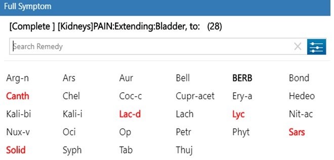
Pain in the kidney region extending to bladder: Berb, Canth, Lyc, Sars, Solid
Pain left-sided: Benz-ac, Coloc, Pareir, Thuj
Pain right-sided: Apis, Lyc, Mag-p, Sars
Pain during urination: Canth, Puls, Senec
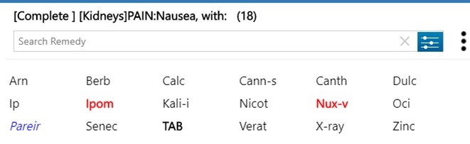
Pain in the kidney with nausea: Canth, Ocimum, Senec
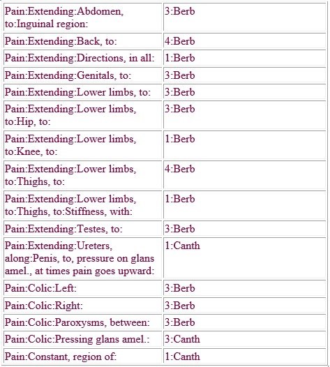
Berberis and Cantharis are the two important homeopathic remedies for kidney stones. Following are the few differentiating rubrics of these two remedies
HOMOEOPATHIC MANAGEMENT
Berberis Vulgaris
Renal colic < left side. Stitching, cutting pain from left kidney following course of ureter into bladder & urethra. Burning & soreness in region of kidneys. Pain in small of back, very sensitive to touch in renal region <when sitting & lying, from jar, fatigue. Numbness, stiffness & lameness with painful pressure in renal & lumbar regions. Bubbling sensation in kidneys. Urine greenish, blood red, with thick, slimy mucus, transparent, reddish or jelly like sediment. Rheumatic & gouty complaints with urinary diseases. < motion, any sudden jarring movement, walking, carriage riding.
Belladonna
Violent spasmodic pains in kidney region especially of the right side. Pain comes suddenly, last indefinitely & cease suddenly. Pains usually in short attacks. Redness of eyes & face, throbbing of brain & carotids. Abdomen tender, distended, < least jar, even of the bed, slight noise, light, lying down. > pressure, tight bandaging, wrapping up. Bilious lymphatic plethoric constitutions.
Cantharides
Constant urging to urinate, passing but a few drops at a time, which is mixed
with blood. Intolerable urging before, during & after urination. Violent
paroxysms of cutting & burning in whole renal region. Violent tenesmus
& strangury. Urine scalds him & is passed drop by drop. Membranous
scales looking like bran in water. Urine jelly like, shredy. Pain raw, sore,
burning in every part, internally & externally. Over sensitiveness of all
parts. Drinking even small quantities of water increases pain in bladder.
Colocynth
Pains on urinating over whole abdomen. Vesical catarrh, discharge like fresh
white of egg. Red hard crystals. Renal colic < left side. Agonising pain in
abdomen causing patient to bend double, with restlessness, twisting &
turning to obtain relief. > hard pressure. Pains < eating & drinking >
warm application. Shooting pains like electric shocks. Complaints from anger,
indignation, mortification.
Dioscorea
Renal colic with pains radiating to the extremities. Colic pains < bending forward & while lying. > on standing erect or bending backwards. Violent twisting colic, occurring in regular paroxysms as if abdomen were grasped & twisted by a powerful hand. Pain suddenly shift to different parts, appear in remote localities as fingers & toes.
Lycopodium
Renal colic, right sided. Pain shooting across lower abdomen from right to
left. Pain in back relieved by urinating. Urine slow in coming, must strain.
Retension. Polyuria during night. Red sand in urine. Uric acid diathesis. Child
cries before urinating. Pains drawing, aching < 4-8 pm. Upper part of the
body emaciated, lower part semidropsical. Ailments from fright, anger,
mortification, reserved displeasure. Avaricious, greedy, miserly, malicious,
pussilanimous. Excessive accumulation of flatulence, lower abdomen. > warm
food & drinks.
Nux vomica
Renal colic, right sided. Pain extends to the right thigh & to the genitals. Frequent ineffectual urge for urination with dribbling of urine. Haematuria, strangury. While urinating, itching in urethra & pain in neck of bladder. Backache, must sit up or turn over in bed. Adapted to thin, irritable, zealous, nervous, literary, studious, responsible persons. Bad effects of coffee, tobacco, alcohol, highly spiced food, overeating, long continued mental exertion. Over sensitiveness to all external impressions. Frequent ineffectual urging for stool.
Ocimum canum
Renal colic, right sided. Uric acid diathesis. Red sand in urine. High acidity, formation of spike crystals of uric acid. Turbid, thick, purulent, bloody, brick dust red or yellow sediment. Odour of musk. Pain in ureters, cramps in kidneys.
Pareira brava
Renal colic, pain going down the thighs. Neuralgic pain in the anterior crural region. Constant urging, great straining. Can emit urine only when he goes on his knees, pressing head firmly against floor. Black, bloody, thick mucus urine. Dribbling after micturition. Urethritis, prostatitis.
Sarsaparilla
Passage of small calculi or gravel, renal colic, stone in the bladder. Excruciating pains from right kidney downwards. Severe almost unbearable pain at conclusion of urination. Urine bloody, scanty, slimy, flaky, sandy, copious, passed without sensation, deposits white sand. Painful distension & tenderness in bladder, urine dribbles while sitting, passes freely when standing. Air passes from urethra, child screams before & while passing urine.
Solidago
Urine scanty, reddish brown, thick sediment, dysuria, gravel. Pain in kidneys extends forward to abdomen & bladder. Urine difficult & scanty, albumen, blood & slime. Kidneys sensitive to pressure. Backache of congested kidneys.
REFERENCES
- Young VB, Kormos WA, Chick DA et-al. Blueprints Medicine. Lippincott Williams & Wilkins. (2009) ISBN:0781788706. Read it at Google Books – Find it at Amazon
- Elsevier saunders kumar and Clarke clinical medicine, 6th edition by Praveen kumar and Michael Clarke.
- https://books.google.co.in/books/about/Clinical_Medicine.html?id=xlEwZWFVFDcC
- Zomeo Homeopathic Software; https://hompath.com/free-homeopathic-software
- Allen.H.C, Allen’s keynotes rearranged and classified with leading remedies of the materia medica and bowel nosodes, 10 th edition,Newdelhi, B.Jain publishers.
- Boericke William , Pocket manual of homoeopathic materiamedica and repertory comprising the characteristics and guiding symptoms of all remedies (clinical and pathogenetic) including Indian drugs, 9 th edition, Newdelhi, B.Jain publishers.