ABSTRACT
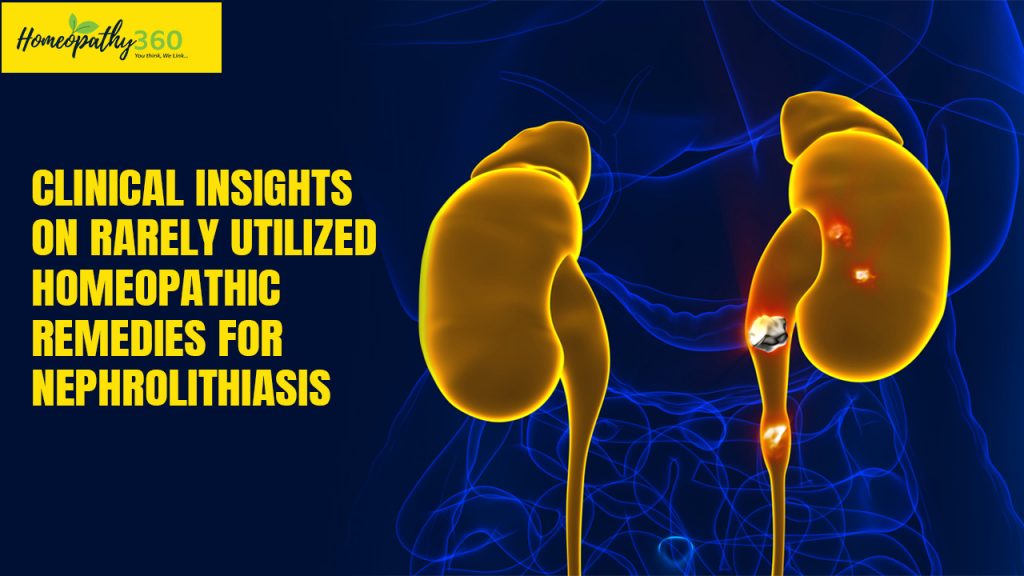
Nephrolithiasis is the formation of urinary calculi in the kidney, which may be deposited along the entire urogenital tract. Urinary stones can be composed of calcium, uric acid, struvite (due to infection), or cystine. Nephrolithiasis manifests as an acute, sudden, sharp and wavy pain in the back and its whole side, which can be moved to the lower abdomen or genital space. This article discusses the role of less commonly known rare homeopathic medicines for nephrolithiasis.
KEYWORDS
Nephrolithiasis, Rare homeopathic remedies, Cystine stones, Calcium stones, Uric acid stones, Infection stones
INTRODUCTION
‘Nephrolithiasis’ is a term used to describe the presence of urolithic crystals, sometimes known as stones, in the urinary tract. The annual incidence of urolithiasis in the west is around 0.5%, and the lifetime risk of having it is about 10-15%, but in the middle east, it is rising by 20-25%. These calculi/stones are produced through the deposition of polycrystalline aggregates made up of varying concentrations of crystalloid and organic matrix.6
CLASSIFICATION BASED ON COMPOSITION OF STONE
Calcium stones
The second part of the 18th century saw the discovery of oxalate and uric acid stones by Karl W. Scheele (1742-1786). Both the monohydrate (also known as whewellite) and the dihydrate (also known as weddellite) forms of calcium oxalate stones (CaOx) are possible. This distinction is crucial because dihydrate stones are linked to hypercalciuric conditions and monohydrate stones to hyperoxaluric conditions in the underlying diseases. Idiopathic CaOx stone formers are the great majority of CaOx stone sufferers; they lack any underlying systemic diseases.2
Uric acid and urate stones
Scheele discovered that uric acid was a common component of stones. When the pH of the urine is abnormally low, uric acid stones can form. The other primary factor that leads to uric acid stones is hyperuricosuria. Healthy people have an average daily total urine urate concentration of 500 mg (3 mmol/day). Only 180 mg/L (1 mmol/L) of the total urate species may dissolve at a pH of 5.35. Low ammonia excretion, as well as other circumstances that cause an excessive acid load or alkali loss, including persistent diarrhoea, can all contribute to low urine pH. Patients with gout, diabetes mellitus, metabolic syndrome, and a high protein diet are more likely to have low urine pH and uric acid stones.2
Infection stone
When there is a recurring infection with urease-positive microorganisms including Proteus and Providentia species, as well as some strains of Klebsiella pneumoniae and Serratia Marcescens, magnesium ammonium phosphate stones (also known as struvite), can develop.2
Typically, this group considers stones to be struvite-containing and the outcome of a long-lasting urinary tract infection (UTI) caused by urea-splitting organisms such proteus, staphylococcus epidermidis, urea plasma urea lyticum, and others. Escherichia coli and other bacteria have the ability to cause Calcium Phosphate to precipitate, according to some researchers.3
Cystine stones
Wollaston initially recognized the first cystine stone in 1810 and named it cystic oxide. Since urinary cystine is typically not routinely measured at the first kidney stone presentation, therefore the diagnosis of cystine stones depends on a high index of suspicion. Young age of presentation, mildly radiopaque stones, family history, and intraoperatively sulphate odor from stone fragmentation are factors that should raise suspicion. To make the diagnosis, a 24-hour urine test or stone analysis is required.2
CLINICAL FEATURES6
- An acute, sudden, sharp and wavy pain in the back and its whole side, which can be moved to the lower abdomen or genital space.
- A feeling of sudden urination.
- Burning feeling at urination.
- The color of the urine will be dark or red due to blood particles of RBCs.
- Feeling of nausea and vomiting.
- Male patients feel pain at the tip of their penis.
RISK FACTORS 4
Low fluid intake
Low fluid intake is the single biggest factor in stone formation. A low fluid intake leads to concentrated urine production, which super saturate and crystallizes compounds that cause stones. Low urine flow rates also encourage the deposition of crystals on the urothelium.
Hypercalciuria
The most prevalent urine chemistry abnormality in people who develop stones repeatedly is high urinary calcium levels. The finding that the most consistent difference between stone-formers with and without a family history of stones is high urine calcium suggests that “idiopathic” hypercalciuria has a genetic basis.
Deactivating VDR variants
Patients with deactivating VDR mutations develop stones if hypocitraturia is present. Citrate excretion is encouraged by VDR activity, which raises the solubility of calcium salts. In these patients, a diet lacking in fruits and vegetables may increase citrate excretion, which increases the risk of stone formation. Supplementing with citrate might be a particularly successful treatment strategy.
Primary hyperparathyroidism
In primary hyperparathyroidism, activated extracellular calcium–sensing receptor (CaSR) causes urinary dilution and acidification, protecting against stone formation by preventing supersaturation of urine with insoluble calcium salts. Stones develop in hyperparathyroid patients with allelic variants that cause reduced expression of the CaSR, with associated loss of its urinary dilution and acidification effect.
High salt diet
A high-sodium diet raises the calcium production of the urine. The electric gradient necessary for paracellular calcium absorption is driven by the sodium/chloride co-transporter at the Henle loop. One element of a successful strategy to lower nephrolithiasis rates in one trial was a lowered salt intake.
INVESTIGATIONS1
- Plain X-ray KUB – For radio-opaque calculations. 90% of stones may be diagnosed with it. Additionally, a larger renal shadow is evident.
- CT scan It will locate the little stones that were overlooked for non – opaque calculi.
- USG – Its location, size, and confirmation of the kidney’s enlargement make it the most valuable stone diagnostic.
- Blood Examination – creatinine, serum calcium, and serum uric acid
HOMOEOPATHIC MANAGEMENT7
Stigmata maydis-Zea maydis: Used when there is insufficient micturition. Phosphatic and uric diathesis. Cystitis. urine retention and suppression. Dysuria. Renal lithiasis, nephritic colic, and blood and red sand in the urine. After micturating, tenesmus. Vesical catarrh. Cystitis.
Oxydendron arboreum- Andromeda arborea (sorrel tree): Obstructed urination. Deranged portal circulation. Enlargement of the prostate. Vesical calculi. Discomfort in the bladder neck.
Ipomoea bona-nox: Stooping causes pain in the left lumbar muscles. Back pain associated with kidney diseases. Aching in the lower back and extremities; renal colic.
Xanthorrhoea arborea: Renal pain that is unbearable, gravel, and cystitis. Testicles, bladder, and ureters all experience pain; the slightest amount of cold or dampness causes lumbar pain
Coccus Cacti: Urine contains albumin. Urine sediment is dark red. Pain that radiates from the kidney to the bladder and down the legs.
Epigea Repens: Dysuria, chronic cystitis, tenesmus after urination, muco-pus and uric acid deposit, gravel, and renal calculi are some of the symptoms. Brown colored fine sand in the urine. Tenesmus and burning in the bladder neck after urinating.
Piperazinum: Uric acid diathesis. Gout and calculi in the urine. Persistent back pain. Urine scanty and skin dry.
Galium Aparine: It works as a diuretic on the urinary organs and is useful for calculi, dropsies, and gravel. Cystitis and dysuria.
Barosma Crenulatum: Mucopurulent discharges; markedly particular effect on the genito-urinary system. Bladder irritation, including cystitis; prostatic conditions. Gravel.
CONCLUSION
Homeopathy saves the individual from invasive procedures hence it is a safer option for those seeking treatment for nephrolithiasis. The medicines are given in minute doses and derived from plants, minerals, and animals. Homeopathy treats the cause of the disease, therefore reduces the risk of recurrence. Hence it is a better choice for individuals looking for treatment of nephrolithiasis.
REFERENCES
- Singh V. Renal Calculi and its Homoeopathic approach. International Journal of Homoeopathic Sciences 2021; 5(2): 84-86
- Viljoen A, Chaudhry R, Bycroft J. Renal stones. Annals of clinical biochemistry. 2019 Jan;56(1):15-27.
- Daudon M, Bader CA, Jungers P, Beaugendre O, Hoarau MP. Urinary calculi: review of classification methods and correlations with etiology. Scanning Microscopy. 1993;7(3):32.
- Dawson CH, Tomson CR. Kidney stone disease: pathophysiology, investigation and medical treatment. Clinical medicine. 2012 Oct;12(5):467.
- Sohgaura A, Bigoniya P. A review on epidemiology and etiology of renal stone. Am J Drug Discov Dev. 2017 Mar 15;7(2):54-62.
- Khan F, Haider MF, Singh MK, Sharma P, Kumar T, Neda EN. A comprehensive review on kidney stones, its diagnosis and treatment with allopathic and ayurvedic medicines. Urol Nephrol Open Access J. 2019;7(4):69-74.
- Boericke W. New manual of homoeopathic materia medica & repertory with relationship of remedies: Including Indian drugs, nosodes uncommon, rare remedies, mother tinctures, relationship, sides of the body, drug affinities & list of abbreviation: 3rd edition. New Delhi, India: B Jain.
ABOUT THE AUTHORS
Dr. Navroop Bhatia, PG Scholar Part-1, Department of Materia Medica, Dr. MPK Homoeopathic Medical College, Hospital & Research Centre, Jaipur.
Dr. Pooja Patidar, PG Scholar Part-1, Department of Materia Medica, Dr. MPK Homoeopathic Medical College, Hospital & Research Centre, Jaipur.
Dr. Tanya Vaish, PG Scholar Part-1, Department of Materia Medica, Dr. MPK Homoeopathic Medical College, Hospital & Research Centre, Jaipur.
Dr. Sonia Tuteja, Professor, Dr. MPK Homoeopathic Medical College, Hospital & Research Centre, Jaipur.


