SKIN
THE TINEA VERSICOLOR
Oct 31.10.2020 by SHARON.X,BHMS-FINAL YEAR
Introduction:
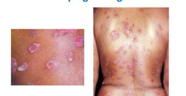
The most common type of skin manifestations in tropical countries is Tinea versicolor.This occurs due to fungal infection of the skin.The fungus interferes with the normal pigmentation of the skin, resulting in small, discoloured patches. These patches may be lighter or darker in colour than the surrounding skin and most commonly affect the trunk and shoulders. Sometimes the patches may also occur in face, which especially in young girls they feel stressed due to their irregular skin colour.
KEYWORDS:Skin,lighter or darker skin patches,fungal infection, homoeopathic approch
Firstly lets have a precise view on anatomy and physiology of skin; The skin is the largest organ of the body, accounting for about 15% of the total adult body weight. It performs many vital functions, including protection against external physical, chemical, and biologic assailants, as well as prevention of excess water loss from the body and a role in thermoregulation. The skin is continuous, with the mucous membranes lining the body’s surface. The skin is composed of three layers: the epidermis, the dermis, and subcutaneous tissue.
OUTER LAYER:
The epidermis- is a stratified; squamous epithelium layer consists of layers of flattened cells.
The epidermis has three main types of cell:
• Keratinocytes (skin cells)
• Melanocytes (pigment-producing cells)
• Langerhans cells (immune cells).
• The Merkel cell is a fourth, less visible, epidermal cell. Their exact role and function are not well understood.
MIDDLE LAYER:
The dermis- is the fibrous connective tissue or supportive layer of the skin. The major fibres are:
• Collagen fibres: These have enormous tensile strength and provide the skin with strength and toughness. Collagen bundles are small in the upper or papillary dermis and form thicker bundles in the deeper or reticular dermis.
• Elastin: this type of fibre provides the properties of elasticity and pliability to the skin.
The normal cells in the dermis include:
• Mast cells-These contain granules packed with histamine.
• Vascular smooth muscle cells-These allow blood vessels to contract and dilate, required to control body temperature.
• Specialised muscle cells-For example, myoepithelial cells are found around sweat glands and contract to expel sweat.
• Fibroblasts-These are cells that produce and deposit collagen and other elements of the dermis as required for growth or to repair wounds.
• Immune cells- The role of tissue macrophages (histiocytes) is the phagocytosis.There are also small numbers of lymphocytes in the normal dermis.
INNER LAYER:
The subcutis -is the fat layer immediately below the dermis and epidermis. The subcutis mainly consists of fat cells (adipocytes), nerves and blood vessels. Fat cells are organised into lobules, which are separated by structures called septae. The septae contain nerves, larger blood vessels, fibrous tissue and fibroblasts. Fibrous septae may form dimples in the skin (so-called cellulite).
ORGANISM:MALASSEZIA
Pityriasis versicolor is caused by mycelial growth of fungi of the genus Malassezia.They are dependent on lipid for survival. Fourteen different species of malassezia have been identified. The most common species cultured from pityriasis versicolor are M. globosa, M .restricta and M sympodialis.Usually malassezia grow sparsely in the seborrhoeic areas (scalp, face and chest) without causing a rash.
The yeasts induce enlarged melanosomes (pigment granules) within basal melanocytes in the brown type of pityriasis versicolor. It is easier to demonstrate the yeasts in scrapings taken from this type of pityriasis versicolor than in those taken from the white type.
The white or hypo pigmented type of pityriasis versicolor is thought to be due to a chemical produced by malassezia that diffuses into the epidermis and impairs the function of the melanocytes.
The pink type of pityriasis versicolor is mildly inflamed, due to dermatitis induced by malassezia or its metabolites. Pink pityriasis versicolor and seborrhoeic dermatitis may co-exist, as both are associated with malassezia.
Hyper pigmented, hypo pigmented and inflamed pityriases versicolor are usually seen as distinct variants but may sometimes co-exist.
DIFFERENT TYPES OF TINEA:
• Tinea capitis- fungal infection of the scalp.
• Tinea cruris – fungal infection in the folds of the skin [jock itch]
• Tinea corporis – superficial fungal infection mostly on arms and legs , also known as ringworm or dermatophytosis.
• Tinea pedis – fungal infection usually begin in toes [Athlete’s foot]
• Tinea unguium – fungal infection of the nails [onychomycosis]
• Tinea nigra – fungal infection causing dark or brown painless patches in skin [superficial phaeohyphomycosis]
HOMOEOPATHIC APPROCH:
Top grade homeopathic medicines for tinea versicolor include Arsenic Album, Sepia, Thuja Occidentalis, Sulphur, Tellurium, Natrum mur. ,Psorinum and Kali Sulph. Homeopathic medicines can be given according to the patient’s individual symptoms and this will helps to boost the infection fighting mechanism of the body and help to fight fungal infection.
REFERENCE:
• https://dermnetnz.org/topics/pityriasis-versicolor/
• Allen’s keynotes
• https://www.google.com/search?q=tinea+versicolor&oq=tinea+versicolor&aqs=chrome..69i57.6869j0j8&sourceid=chrome&ie=UTF-8