Scabies and Its Homoeopathic Management
ABSTRACT:
Scabies has been a common companion of the human species for over 2500 years. It affects around 100–300 million people worldwide. Scabies is an intense pruritic disease affecting the skin caused by the ectoparasitic Sarcoptes scabiei var hominis. Transmission is by close contact, infested clothes, overcrowding. Infection of a new host is initiated by the contact with the infected person and the female mite that burrows into the superficial layers of the skin to lay eggs. Intensely itchy papules in the classical sites- interdigital spaces of fingers, genitalia. The diagnosis should be suspected with a clinical history of itch, worse at night, affecting other family members, clinical distribution, and appearance. As it is a contagious disease proper personal hygiene is to be maintained and the clothes and bed linen are disinfected by laundering or hot ironing. Homoeopathy offers an individualized approach for the treatment of Scabies. This article reviews about Scabies, the pathogenesis, clinical features, diagnosis, general management and the homoeopathic therapeutics against it.
KEYWORDS: Scabies, Sarcoptes scabiei var. hominis., Homoeopathy.
INTRODUCTION:
Scabies was first described in the Old Testament and by Aristotle. The name Sarcoptes scabiei is derived from the Greek word “sarx” that means flesh and “koptein” that means to cut, and the Latin word “scabere” that means to scratch.1
Scabies is an ectoparasitic infection caused in humans by the Sarcoptes scabiei var. hominis.2
DESCRIPTION:2
The adult female measures approximately 0.4 mm long by 0.3 mm broad, and the smaller male 0.2 mm long by 0.15 mm broad. The body is creamy white and is marked by transverse corrugations, and on its dorsal surface by bristles and spines. There are four pairs of short legs; the anterior two pairs end in elongated peduncles tipped with small suckers. In the female, the rear two pairs of legs end in long bristles (setae), whereas in the male bristles are present on the third pair and peduncles with suckers on the fourth.
Copulation occurs in a small burrow excavated by the female. The burrow is not confined to the stratum corneum, but is inclined downwards into the epidermis. Approximately 40–50 eggs are laid by each female during a lifespan of 4–6 weeks. Eggs hatch after 3–4 days into larvae, which dig new burrows closer to the skin surface. There, the larvae mature into adult mites in about 4 days.
Adult females can live in the host for up to a month. The life cycle lasts around 14–21 days. The mites show a preference for certain sites in which to burrow, and appear to avoid areas with a high density of pilosebaceous follicles. Approximately 40–50 eggs are laid by each female during a lifespan of 4–6 weeks. Away from the host, scabies mites survive for 24–36h at room conditions (21°C and 40–80% relative humidity).
EPIDEMIOLOGY:2
Scabies is a worldwide problem and all ages, races and socioeconomic groups are susceptible. Scabies affects around 100–300 million people worldwide, but accurate figures of its prevalence are difficult to obtain.
Crusted scabies (formerly called Norwegian scabies) is found in individuals with compromised immune systems (e.g., the elderly, people infected with HIV, and transplant patients) as well as those with decreased sensory functions (e.g., patients with leprosy or paraplegia). Such individuals experience minimal pruritus despite their highly contagious state
Age: Prevalence is higher in children and in people who are sexually active. Scabies occurs in all age groups. However, it becomes frequent in the elderly in residential and nursing home environments.
Sex ratio: The overall sex incidence is probably equal.
Ethnicity: Whereas all ethnic groups are susceptible, there are some differences which are probably related to customs and social factors rather than inherent susceptibility.
Environmental factors: Hastens the spread include overcrowding, delayed treatment of primary cases, and lack of public awareness of the condition. Higher incidences occur with overcrowding, often related to natural disasters, wars, economic depression and refugee camps.
Scabies can be transmitted directly by close personal contact, sexual or otherwise, or indirectly via fomite transmission. Spread of the infestation between family members and close contacts is common.
PATHOGENESIS:1,2,3
Scabies is usually transmitted by close physical contact, such as prolonged hand‐holding or the sharing of a bed.
The incubation period before symptoms develop can range from days to months. In first-time infestations, it usually takes 2–6 weeks before the host’s immune system becomes sensitized to the mite or its by products, resulting in pruritus and cutaneous lesions. A subsequent infestation is usually recognized within 24–48 hours. Asymptomatic scabies-infested individuals are not uncommon, and these individuals can be considered ‘carriers.’
It has been estimated that a patient with conventional scabies needs between 15 and 20 minutes of close contact to transfer the mites from one person to another. The average patient can have between 5 and 12 mites, and it is higher in crusted scabies, where the patient can have millions of mites, therefore patients are more contagious especially for the medical and paramedical staff.
Host immune response: Once the S. scabiei penetrates the skin, it releases substances in response to contact with keratinocytes and Langerhans cells, initiating an inflammatory and immune reaction that involves multiple cell lines.
The reaction consists of both type I and type IV hypersensitivity reactions.
In the type I reaction, an antigen on the mite encounters specific immunoglobulin-E (IgE) on mast cells within the epidermis, leading to the degranulation of mast cells causing wheal-and-flare reactions.
In the type IV hypersensitivity reaction, patients that have not had contact with the mite will have between 10 and 30 days before the rash is noticeable. When patients are infected for a second time, the hypersensitivity reaction may develop within a day.
Scabies exhibits strong superficial and deep dermal and perivascular inflammatory infiltrates composed of lymphocytes, histiocytes, and eosinophils, and will eventually have elevated antibody titers specific for antigens from the parasite. There is marked increase in the secretion of interleukin 6 (IL-6) and vascular endothelial growth factor (VEGF), and slight increase in secretion of granulocyte colony stimulating factor (G-CSF) from normal human epidermal keratinocytes.
IL-6 is known to stimulate the proliferation of keratinocytes; IL-8 and G-CSF promote monocytes becoming dendritic cells and the proliferation of neutrophils.
IL-6 is also known to activate Th1 CD4+ cells to secrete IL-2, promoting their proliferation and differentiation, to activate Th2 CD4+ cells to produce IL-4, which drives antibody production known to increase vascular permeability, initiating inflammation, causing the edema in scabies lesions.
Molecular studies: Data from S. scabiei complementary DNA (cDNA) libraries have identified several unique genes. Among the cDNAs encoding for S. scabiei are homologues of the known house dust mite allergens glutathione-S transferase, paramyosin, and cathepsin-L.
PATHOLOGY:3
A patchy to diffuse infiltrate with eosinophils is noted in the reticular dermis. On transection, a scabies mite may occasionally be seen within the epidermis. Pink ‘pigtail’-like structures attached to the stratum corneum (which represent fragments of the adult mite exoskeleton) can serve as a clue to the diagnosis of scabies when mites, scybala or eggs are not identified.
CLINICAL PRESENTATION:1,2,3
- The epidemiologic history (e.g., pruritus in household members or close personal contacts), the distribution and types of lesions, and pruritus form the basis of the clinical diagnosis.
- Itching is the most obvious manifestation of scabies, usually sparing the face in adult classic scabies. The intense pruritus classically is accentuated at night and is exacerbated by a hot bath or shower. These symptoms may be present before any overt physical signs appear.
- The onset occurs 3–4 weeks after the infection is acquired.
- Reinfection of a previously cured individual, however, may provoke immediate symptoms.
- Cutaneous lesions are symmetrical.
- Typical sites of involvement include the interdigital webbing of the hands, the flexural aspect of the wrists, axillae, behind the ears, waist, ankles, feet, buttocks and belt area.
- In men, penile and scrotal lesions are common, while in women, the areolae, nipples and genital area are often affected.
- In infants, the elderly and immunocompromised hosts, all skin surfaces are susceptible, including the scalp and face.
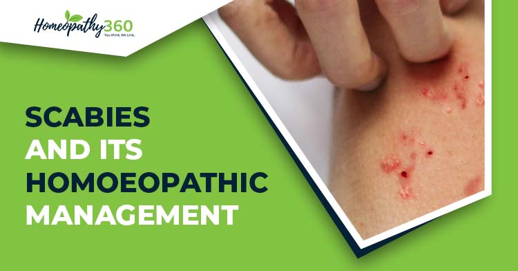
The typical lesions of scabies are burrows, which appear as slightly raised brownish tortuous lesions.
- Nodules are intensely itchy, and may persist for weeks or months after the scabies has been effectively treated. Secondary lesions are not specific.
- They include excoriations, eczematization and impetiginization, and may occur anywhere.
- Commonly, small erythematous papules are present, associated with variable numbers of excoriations.
- The pathognomonic sign is the burrow, representing the tunnel that a female mite excavates while laying eggs.
- Clinically, the burrow is wavy, thread-like, greyish-white and 1–10 mm in length.
- Many patients, however, do not have any obvious burrows on inspection, especially in warm climates.
INVESTIGATIONS:2,3
- The typical history of pruritus with nocturnal exacerbations, the presence of contact cases within the family and the distribution of the eruption of inflammatory papules, should suggest the diagnosis.
- Failure to find mites is common and does not rule out scabies.
- With mineral oil examination in which skin scrapings from infested areas are inspected under light microscopy for adult mites, eggs and/or fecal pellets. A scalpel or curette may be used to obtain the skin sample. At low magnification, the circumflex accent‐like image (as the French letter ‘ô’) represents the head and the two pairs of front legs of the mite.
- In some patients, epiluminescence microscopy or dermoscopy can prove useful for direct in vivo visualization of mites and eggs.
- A skin biopsy may confirm the diagnosis of scabies if a mite or parts can be identified.
- However, in most cases, the histology shows non‐specific features, with epidermal spongiosis, papillary oedema, and superficial and deep perivascular inflammatory cell infiltrates with numerous eosinophils
- Adhesive tape test may be useful in particular situations. After firmly applying the adhesive side of the tape onto an appropriate skin lesion of patients, the tape is pulled off and transferred directly onto a slide for microscopy, affixing the adhered separated part of the corneal skin. however, its sensitivity is low.
In the absence of confirmed mites, diagnosis is currently based entirely on clinical and epidemiological findings.
DIFFERENTIAL DIAGNOSIS:3
Unless skin burrows are noted clinically, all diseases associated with pruritus and infected lesions have to be considered in the differential diagnosis. In particular, atopic, contact or nummular dermatitis, autosensitization (‘id’ reaction), pyoderma, dermatitis herpetiformis, bullous pemphigoid, and other insect bites should be considered.
GENERAL MANAGEMENT: INDICATION FOR THERAPY2
Treatment should be prescribed to the patient and close physical contacts, even without pruritus or cutaneous lesions.
Patients should be advised to avoid close physical contact until they and their household members and sexual partners have been treated. A detailed verbal and written information about scabies infestation should be given to the patient.
HOMEOPATHIC MANAGEMENT:
MURPHY- HOMOEOPATHIC MEDICAL REPERTORY:5
Clinical: SCABIES, general-
agn., aloe,alum., ambr., ant-c., ant-t., anthro., apis, ARS, aster., bar-m., bell., bry., calc., canth., carb-ac.,carb-an., CARB-V., CARBN-S., CAUST., chin., clem., coloc., con., cop., croto-t., cupr., dulc., elaps., ferr-ma., graph., guai., hep., iod., KALI-S., kreos., lach., led., lyc., m-ambo., mang., merc., merc-i-f., mez., nat-c., nat-m., nat-s., nit-ac., nux-v., olnd., petr., ph-ac., PSOR., puls.,rhus-t., rhus-v.,rumx., sabad., sarr., sars., SEL., SEP., sil., squil.,staph., sul-ac., SULPH.,tarax., thuj., valer., verat., zinc.
FEW HOMOEOPATHIC THERAPEUTICS ON SCABIES:6,7,8
Arsenicum Album: Desquamation of the skin of the body. The skin is dry as parchment, hot itching, and violent burning in the skin. Vesicular itchy eruptions which violently burn, esp. at night, or with coverings, Fetid smell, ichorous suppuration, ready bleeding, putridity, Discoloured itching, burning, swellings; edema, eruption, papular, dry, rough, scaly.
Carbo Vegetabilis: Eruption of small pimples like miliary scabies, streaks of a reddish brown. Itching; worse on evening, when warm in bed. Moist skin; hot perspiration; burning pain. Ichorous, offensive discharge, sensation of tingling of the skin, throughout the body. Burning sensation in different parts of the skin. Painless ulcers in the extremities of the fingers and of the toes. Fetid ulcers, with burning pains, and discharge of corrosive and bloody pus.
Carboneum Sulphuratum: Furunculosis. Chronic skin diseases with much itching. Biting after scratching. Burning of the skin after scratching. Dryness with burning. Eruptions. Biting; blisters; boils; burning; discharges, corrosive, glutinous, yellow fluid; dry; eczema. Herpetic eruptions; scabby; scaly; tearing pain. Eruptions; itching; like measles; phagedenic; pimples; pustules; rash after scratching. Scabby eruptions, moist, worse after scratching. Smarting eruptions. Eruptions worse after scratching. Vesicles filled with yellow fluid. Erysipelas with much swelling and covered with vesicles. EXCORIATION; after scratching. Formication all over body. Indurations in skin. ITCHING AT NIGHT; in a warm bed. Sticking in the skin; after scratching. Ulcerative pains in skin.
Causticum: Soreness in folds of skin, back of ears, between thighs. Violent burning itching, esp. in the back, and in the calves of the legs. Itching of the whole body at night. Tingling (or stinging) swelling (sometimes called “buzzing” swelling). Miliary eruptions and nettle-rash. Itching and humid tetters. Ulcerative vesicles, burning ulcers, with yellowish-looking skin; ulcers burning, with corroding pus, with thin or watery pus, suppurating; jerking pains running through the ulcers. Excoriation in children.
Graphites: Eruptions, oozing out a sticky thick honey-like exudation. Cracks in nipples, mouth, between toes, anus. Phlegmonous erysipelas of face; burning and stinging pain. Obstinate dryness of the skin, and absence of perspiration. Red spots on the skin, like flea-bites. Erysipelatous inflammations. Vesicular erysipelas, on the abdomen and on the back. Tetters, and other humid or scabby eruptions, sometimes with secretion of corrosive serum, or with itching in the evening, and at night. Eruption of pimples and nodules (principally under hair and on covered parts) which itch very much. Excoriation of the skin (in the bends of the limbs, groins, neck, behind the ears), esp. in children. Ulcers discharging a glutinous fluid, thin and sticky.
Kali sulphuricum: Burning, itching, papular eruption exuding pus-like moisture. Fine red pimples running together. Scurf, scaling, chapping, sores with yellow, sticky secretions. Sensation of burning, in the skin; burning after scratching. Eruptions, blisters, burning, dry, moist; eczema with yellowish green, watery, discharge, herpetic. Itching and stinging eruptions. Rash like measles. Eruptions painful, pimples, psoriasis, pustules, red eruptions. Scabby, vesicular eruptions. Erysipelas with blisters. Easy excoriation of the skin. Stinging; aggravated when warm in bed; ameliorated scratching. Moisture of the skin after scratching.
Psorinum: Intolerable itching. Herpetic eruptions, especially on scalp and bends of joints with itching; worse, from warmth of bed. Crusty eruptions all over, pustules near finger-nails. Pimples: on forehead; on neck and mammae; with black points in centre, painful when scratched; on external throat. Boils on chest and loins; on buttocks, with burning itching, soon disappearing, leaving crusts. Itch-like eruption on face, hand, back, and leg, and agglutination of eyes. Vesicles: on face; quickly filling with yellow lymph, sore to touch on forehead, face, and behind right ear; filled with lymph, painful to touch on various parts, some forming itching papules. Tickling and heat; of face, neck, and hands on touch; over whole body after rubbing papules and vesicles; between fingers, and vesicles filled with lymph, where a flea had bitten, with white, hard blisters on a red base.
Selenium: Dry, scaly eruption in palms, with itching, Itching about the ankles and folds of skin, between fingers. Itching about finger-joints and between fingers; in palms. Vesicular eruption between fingers. Frequent tingling in circumscribed parts of skin, with great provocation to scratch. Miliary eruption. Prolonged oozing from parts which have been scratched, Itching in folds of skin, between fingers and about joints, esp. ankle joint.
Sepia: Itching; not relieved by scratching; worse in bends of elbows and knees. Soreness of skin and humid places in bends of joints. Itching in different parts (face, arms, hands, back, hips, abdomen and genitals) which changes to a burning sensation. Itching and eruption of pimples in the joints. Excoriation, esp. in the joints. Dry and itching eruptions, like scabies. Dry itch; moist, scabious herpes, with itching and burning sensation. Eruptions of vesicles, like pemphigus. Itching, stinging, lancinating, burning, or sometimes indolent ulcers (knuckles, finger-joints, tips of fingers, joints and tip of toes).
Sulphur: Colicky babies with pimples, itch, or eruption on skin, or roughness of skin. Exanthema in general on any part of the body which is < by any heat, from getting warm at work, in bed, dry; rough; scaly; voluptuous itching “feels so good to scratch”. Tetters in general; chapped; scurfy; painful; tearing; pulsating. Itching in skin, even of whole body, < at night, or in morning, in bed, and often with pain as of excoriation, heat, itching (soreness), or bleeding of the part which has been scratched. Scabious eruptions and tetters of a greenish yellow colour, commencing with small itching phlycten, with a red areola. Miliary eruptions, principally on limbs. Burning itching of the eruptions. Erysipelatous inflammation, with pulsative and shooting pains. Tingling in the skin throughout the body. Excoriation, especially in folds.
COMPLICATIONS:2
Eczematous changes are common, and may be widespread and severe. The inappropriate use of topical steroids may further modify the clinical picture to mimic other dermatoses. Secondary infection, manifest as folliculitis or impetigo, may also be severe and extensive. In the tropics and subtropics, where nephritogenic strains of β‐haemolytic streptococci may be responsible for secondary impetigo, haematuria‐related glomerulonephritis occurs as a complication of scabies.
CONCLUSION:
Scabies is a worldwide problem affecting all age groups. As it can infect one person with another, proper hygiene is to be maintained and disinfecting the clothes properly is required so as to limit the spread. Proper teaching to the community to prevent its spread is also much needed.
ACKNOWLEDGMENT
I heartfully thank my PG Guide, Dr Praveen
Kumar P.D., Professor, HOD and PG Guide of Department of Practice of Medicine, Government Homoeopathic
Medical College Bengaluru, for his valuable guidance.
REFERENCES:
- Hicks MI, Elston DM. Scabies. Dermatologic therapy. 2009 Jul;22(4):279-92. https://www.praxis-schuster.ch/wp-content/uploads/2016/10/Hicks.pdf
- Griffiths C, Barker J, Bleiker T, Chalmers R, Creamer D. Rook’s Textbook of Dermatology. 9th ed. Chichester, West Sussex: John Wiley & Sons Inc.; 2016. 34.39-34.44.
- Bolognia J, Jorizzo J, Rapini R, Schaffer J. Dermatology. 2nd ed. London: Mosby Elsevier; 2008.pg 1291-1293.
- Das K. Textbook of Medicine. 5th ed. New Delhi: Jaypee Brothers Medical Publishers:2008. p1357,1358.
- Murphy R. Homoeopathic Medical Repertory. Revised 3rd ed. Noida: B Jain Publishers (p) ltd; 2010: p463.
- Boericke W. Boericke’s new manual of Homoeopathic Materia Medica and repertory. 3rd revised and augmented ed. New Delhi: B Jain Publishers (P) LTD; 2016.
- Clarke J H. A Dictionary of Practical Materia Medica. Jain Publishers, New Delhi,1982.
- Kent JT. Lectures on Homoeopathic Materia Medica; Reprint Edition: 2006. New Delhi: B Jain Publishers (P) Ltd; 2006.
Author’s Detail:
Dr. Bianghunlang Nongsiej B.H.M.S.
PG Scholar
Department of Practice of Medicine
Government Homoeopathic Medical College Bengaluru, Karnataka-560079
Email: [email protected]
Under the guidance of
Dr Praveen Kumar P.D.M.D.(HOM)
Professor, HOD and PG Guide
Department of Practice of Medicine
Government Homoeopathic Medical College Bengaluru