ABSTRACT
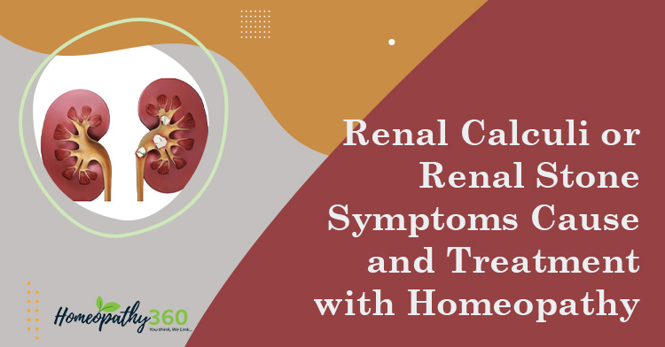
Kidney stones are hard deposits that form in the urine, made up of mineral and acid salts. Kidney stones can be transported throughout the urinary tract and can lodge in the ureters, bladder and urethra. Here the various types of calculi are being described along with their homoeopathic management. This will enhance our studies about the various renal calculi, its therapeutics and scope in homoeopathy.
KEYWORDS
renal calculi, homoeopathy
Abbreviations
Eg – example, ESR – erythrocyte sedimentation rate, KUB – kidney, ureter, bladder, PTH – parathyroid hormone, CT – computed tomography, ESWL – extracorporeal shock wave lithotripsy
INTRODUCTION
Renal calculi is a challenging clinical problem in today’s world. They are small hard, deposits that form in the urine that are made up of mineral and acid salts. Though kidney stones form in the kidneys, they can be transported throughout the urinary tract, which includes the kidneys, ureters, bladder and urethra. In surgery, only the stones are removed not the process of formation of stones so, in such case, homoeopathy comes into play. The prevalence of renal calculi is higher in those living in mountains, desert and tropical areas. Generally, men are more affected than women.1
AETIOLOGY
Deficiency of vitamin A causes desquamation of epithelium.
Dehydration increases the concentration of urinary solutes until they are liable to precipitate.
Decreased urinary citrate
The presence of citrate in urine, as citric acid, tends to keep insoluble calcium phosphate and citrate in solution.
Renal infection
Infection favours the formation of urinary calculi. Clinical and experimental stone formation are common when urine is infected with urea-splitting streptococci, staphylococci, and especially proteus spp.
Inadequate urinary drainage and urinary stasis.
Stones are liable to form when urine does not pass freely.
Prolonged immobilisation
Immobilisation from any cause, e.g., paraplegia, is liable to result in skeletal decalcification and an increase in urinary calcium favoring the formation of calcium phosphate calculi.
Hyperparathyroidism
Hyperparathyroidism leading to hypercalcaemia and hypercalciuria is found in 5% or less of those who present with radio-opaque calculi. Hyperparathyroidism results in a great increase in the elimination of calcium in the urine.²
TYPES OF RENAL CALCULUS
Oxalate calculus (calcium oxalate).
Oxalate stones are irregular in shape and covered with sharp projections, which tend to cause bleeding. A calcium oxalate monohydrate stone is hard and radio dense.
Phosphate calculus
A phosphate calculus [calcium phosphate often with ammonium magnesium phosphate (struvite)] is smooth and dirty white. It tends to grow in alkaline urine. As a result, the calculus may enlarge to fill most of the collecting system, forming a staghorn calculus. Even a very large staghorn calculus may be clinically silent for years until it signals its presence by hematuria, urinary infection or renal failure.
Uric acid and urate calculi
These are hard, smooth and often multiple in number. Pure uric acid stones are radiolucent and appear on an excretion urogram as a filling defect. Most uric acid stones contain some calcium, so they cast a faint radiological shadow. In children, mixed stones of ammonium and sodium urate are sometimes found. They are yellow, soft and friable.
Cystine calculus
These stones appear in the urinary tract of patients with a congenital error of metabolism that leads to cystinuria. Hexagonal, translucent, white crystals of cystine appear only in acid urine. They are often multiple and may grow to form a cast of the collecting system. Cystine stones are radio opaque because they contain Sulphur, and they are very hard.
Xanthine calculus
These are extremely rare. They are smooth and round, brick-red in colour, and show lamellation on cross-section.2
SHAPES OF STONE CRYSTALS IN URINE3
| Types of crystal | Shape of crystal | |
| a. | Calcium oxalate monohydrate | Dumbbell shaped |
| b. | Calcium oxalate dehydrate | Envelope shaped |
| c. | Uric acid | Yellowish of varying size and shape |
| d. | Cystine | Hexagonal, very soft stones |
| e. | Triple stones | Coffin lid shape |
CLINICAL FEATURES
Pain
Silent calculus
Haematuria
Pyuria2
INVESTIGATIONS
Blood
ESR, serum calcium, phosphate, creatinine, blood urea, uric acid, PTH level.3
Radiography
The KUB film shows the kidney, ureters and bladder. An opacity that maintains its position relative to the urinary tract during respiration is likely to be a calculus. Calcified mesenteric nodes and opacities within the alimentary tract can sometimes be shown to be anterior to the vertebral bodies on a lateral radiograph and hence outside the urinary tract.
Contrast-enhanced computerised tomography
CT, preferably spiral, has become the main stay of investigation for acute ureteric colic.
Excretion urography
Urography will establish the presence and anatomical site of a calculus. It also gives some important information about the function of the other kidney.
Ultrasound scanning
Ultrasound scanning is of most value in locating stones for treatment by extracorporeal shock wave lithotripsy (ESWL).2
REPERTORIAL APPROACH
SYNTHESIS REPERTORY
- BLADDER STONES IN KIDNEY
- KIDNEY STONES
- URINE-SEDIMENT
- BLADDER-STONES, bladder, calculi
- KIDNEY STONES, kidney4
MURPHY’S REPERTORY
- Kidneys-STONES, kidney
- URINARY ORGANS-Kidneys-calculi.5
BOGER BOENNINGHAUSEN’S REPERTORY
- URINARY SYSTEM- Urine-sediment, type-Lithic-acid, uric acid, gravel, brick dust.
- URINARY SYSTEM-URINE-TYPE-SEDIMENT-TYPE-Oxalates
- URINARY SYSTEM URINE-TYPE-SEDIMENT-TYPE-Phosphates6
KENT’S REPERTORY
- URINARY ORGANS – BLADDER-CALCULI
- URINE-SEDIMENT7
BOERICKE’S REPERTORY
- URINARY SYSTEM-URINE-TYPE-SEDIMENTS-TYPE Lithic acid, uric acid, gravel, brick dust.
- URINARY SYSTEM-URINE-TYPE-SEDIMENT-TYPE-Phosphates8
HOMOEOPATHIC MANAGEMENT
Berberis vulgaris: Burning pain. Pain in the bladder region. Painful left side from bladder to the urethra. Blood red urine, deposits thick, bright red sediment, slowly becoming clear but always retaining its blood.
Cantharis vesicatoria: Constant and intolerable urging to urinate before, during and after urination. Burning, scalding urine with cutting, intolerable urging and fearful tenses or dribbling strangury. Urine is passed drop by drop. Urine scalds the passage. Jelly like shreddy urine.
Hydrangea arborescens: Burning in the urethra. Urine hard to start. Heavy deposit of mucus. Sharp pain in the loins, especially left. Spasmodic stricture. Profuse deposit of white amorphous salts. Gravel deposits.
Lycopodium clavatum: Renal colic, right sided. Pain shooting across lower abdomen from right to left. Pain in back relieved by urinating. Urine slow in Coming, must strain. Retention. Polyuria during night.
Medorrhinum: Renal colic. Painful tenesmus when urinating. Severe pain in renal region better by profuse urination. Intense pain in ureters, with sensation of passing of calculus. Urine flows very slowly. Aliments from suppressed gonorrhoea.
Ocimum cannuum: High acidity, formation of spike crystals of uric acid. Turbid, thick, purulent (pyuria), bloody (hematuria), brick-dust red or yellow sediment. Odour of musk. Pain in ureters. Cramps in kidney (calculus).
Pareira brava: Black, bloody, thick fucoid urine. Constant urging, great straining, pain down thighs while making efforts to micturate. Can emit urine only when he goes on his knees, pressing the head firmly against the floor. Bladder feels distended. Dribbling after micturition. Violent pain in glans penis. Itching along the urethra.
Sarsaparilla: Passage of gravel or small calculi, renal colic. Stone in bladder, bloody urine. Urine bright and clear but irritating. Scanty, slimy, flaky, sandy, copious, passed without sensation, deposits white sand.
Solidago virgaurea: Scanty, reddish-brown, thick sediment, dysuria, gravel. Difficult and scanty. Albumen, blood and slime in urine. Pain in kidneys extends forward to the abdomen and bladder. Clear and offensive urine. Sometimes makes the use of the catheter unnecessary.
Uva ursi: Calculus inflammation. Chronic vesicle irritation with pain, tenesmus and catarrhal discharge. Burning after the discharge of slimy urine. Frequent urging with severe spasms of the bladder. Urine contains blood, pus and much tenacious mucous with clots in large masses. Painful dysuria. Involuntary, green urine. Cystitis with bloody urine.
Vesicaria: Smarting, burning sensation along urethra and bladder with frequent desire to avoid urine often with strangury. Cystitis, irritable. (8, 9)
Conclusion
Homoeopathy proves to be effective in management of cases of renal calculi.
References
- Thomas S. Professor, Dept. of Repertory, Homoeopathy University, Jaipur. Role of homoeopathy in the treatment of case of ureteric lithiasis with the help of synthesis repertory Journal of Homoeopathy University, Vol.2. April 2016.
- Williams NS, Bullstrode CJ, O’Connell PR. Bailey & Love’s Short Practice of Surgery. Annals of The Royal College of Surgeons of England. 2010 Mar;92(2)
- Bhat S. SRB’s Manual of Surgery. Jaypee Brothers Medical Publishers; 2019 Jun 30.
- Schroyens F, editor. Synthesis: repertorium homeopathicum syntheticum: the source repertory. B. Jain Publishers (P) Limited; 2016.
- Murphy R. Homoeopathic Medical Repertory, A Modern Alphabetical and Practical Repertory. New Delhi (INDIA): B Jain Publication. 2014
- Boger CM. Boenninghausen’s characteristics materia medica & repertory with word index. B. Jain Publishers; 2002.
- Kent JT. Repertory of the homoeopathic materia medica. B. Jain Publishers; 1992.
- Boericke W. Pocket Manual of Homoeopathic Materia Medica & Repertory: Comprising of the Characteristic and Guiding Symptoms of All Remedies (clinical and Pathogenetic [sic]) Including Indian Drugs. B. Jain publishers; 2002.
- Gopi, K., 2022. Kidney Stones – K.S. Gopi. [online] Hpathy.com. Available at: <https://hpathy.com/homeopathy-papers/kidney-stones-2;> [Accessed 24 March 2022].
About the author
Dr Mebanpyntngen Rani
Father Muller Homoeopathic Medical College and Hospital
Deralakatte, Mangaluru 575018
MD Part I Dept. of Homoeopathic Pharmacy


