Abstract: Otorrhea (ear discharge) is a condition frequently met with in clinical and hospital settings, this article briefly describes about this medical condition covering it’s causes, how to deal with the case, making its diagnosis and treatment with special emphasis on some of its good homeopathic remedies.
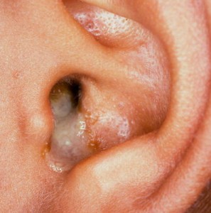
Introduction: Otorrhea is the medical term for ear drainage (discharge from ear). It may be serous, serosanguineous, or purulent. The purulent type of otorrhea is the one most commonly encountered.
Causes:
Otorrhoea may originate from the ear canal, the middle ear, or the cranial vault. Traditionally chronic otitis media is considered to be the commonest cause of otorrhea; however, some studies had proved otitis externa to be the most common cause of otorrhoea. The bacterium most commonly identified in otitis externa is Pseudomonas aeruginosa, Staphylococcus aureus in chronic otitis media and Streptococcus pneumoniae in acute otitis media.
The most common causes of otorrhoea are:
- Otitis externa
- Chronic otitis media (with a perforation of the eardrum, cholesteatoma, or both)
- Acute otitis media with perforation.
The most serious causes are carcinoma of ear and necrotizing external otitis.
Evaluation of Otorrhea:
History of present illness in patients with ear discharge should cover duration of symptoms and whether symptoms have been recurrent. Important associated symptoms include ear pain, itching, decreased hearing, vertigo, pruritus, fever and tinnitus. Patients are questioned about activities that can affect the canal or tympanic membrane (e.g., swimming; insertion of objects, including cotton swabs; use of ear drops), head trauma sufficient to cause a cerebrospinal fluid (CSF) leakage.
Review of systems should seek symptoms of cranial nerve deficit and systemic symptoms suggesting granulomatosis with polyangiitis (e.g., nasal discharge, cough, joint pains).
Past medical history should note any previous known ear disorders, ear surgery (particularly tympanostomy tube placement), and diabetes or immunodeficiency.
Physical examination
Examination begins with a review of vital signs for fever. Ear and surrounding tissues (particularly the area over the mastoid) are inspected for erythema and oedema. The pinna is pulled and the tragus is pushed gently to see whether pain is worsened. The ear canal is inspected with an otoscope; the character of discharge and presence of canal lesions, granulation tissue, or foreign body are noted. Oedema and discharge may block visualization, irrigation should not be used in case if there is a tympanic membrane perforation, but, when possible, the tympanic membrane is inspected for inflammation, perforation, distortion, and signs of cholesteatoma (e.g., canal debris, polypoid mass from tympanic membrane).
When the external ear canal is severely swollen (e.g., as with severe otitis externa) or there is copious drainage, careful suctioning can permit an adequate examination and also allow treatment ( e.g., application of drops, with or without a wick).
The cranial nerves are tested. The nasal mucosa is examined for raised, granular lesions, and the skin is inspected for vasculitic lesions, both of which may suggest granulomatosis with polyangiitis.
Interpretation of findings
Otoscopic examination can usually diagnose perforated tympanic membrane, external otitis media, foreign body, or other uncomplicated sources of otorrhea. Some findings are highly suggestive while other findings are less specific but indicate a more serious problem that involves more than a localized external ear or middle ear disorder:
- Vertigo and tinnitus (disorder of the inner ear)
- Cranial nerve deficits (disorder involving the skull base)
- Erythema and tenderness of ear, surrounding tissues, or both (significant infection)
The following findings are of particular concern:
- Recent major head trauma
- Any cranial nerve dysfunction (including sensorineural hearing loss)
- Fever
- Erythema of ear or periauricular tissue
- Diabetes or immunodeficiency
Differential Diagnosis:
Many cases of ear discharge are clear after clinical evaluation.
If CSF leakage is in question, discharge can be tested for glucose or beta-2 transferrin; these substances are present in CSF but not in other types of discharge.
Malignancy – Patients without an obvious etiology on examination require audiogram and CT of the temporal bone or gadolinium-enhanced MRI. When auditory canal granulation is present, biopsy should be considered if clinical evaluation and CT are not clearly consistent with cholesteatoma.
Treatment:
Treatment is directed at the cause of the ear discharge. Conventional treatment involves use of Antibiotics oral or ear drops, analgesics and antipyretics.
Homoeopathic therapeutics: Natrium muriaticum and Kalium muriaticum work wonders for white coloured ear discharge; Mercurius solubilis and Kalium sulphuricum for yellow; Pulsatilla and Lycopodium for green; Calcarea sulphurica and Mercurius solubilis for blood-stained discharge.
- Kalium muriaticum: Is a top listed medicine for ear discharge. Chronic, catarrhal conditions of the middle ear. Glands about the ear swollen. Snapping and noises in the ear. Threatened mastoid. Great effusion about the auricle.
- Silicea: Is highly recommended for ear discharge with pus. Discharge offensive or fetid odor. Otorrhoea with mastoiditis.
- Psorinum: Works well in chronic otorrhoea when other remedies fail. Otorrhoea offensive, fetid and putrid in nature. The discharge may be brownish in colour and purulent in nature. An intolerable itching, Sensation as if something burst suddenly when eating or swallowing saliva.
- Tellurium: Has cured otorrhoea of long standing in sixth potency; higher dilutions having failed. Discharge fetid, smelling like fish brine stained with blood, thin and watery serum with excoriation and blistering. Intense acridity and excoriating nature of the ear discharge.
- Mercurius solubilis: Works well in cases of ear discharge that are accompanied by an earache. The pain is tearing and shooting in nature. The pain in the ear is worse at night. The discharge from the ear is offensive and mainly yellow in colour. It may also be blood stained. Top listed medicine for swimmer’s ear infection.
- Viola odorata: Shootings in (and around) ears. Had cured discharges of both ears with deafness after one dose of Q. Brings on a discharge that has stopped; or heals a discharge in a few days.
- Kalium bichromium: Stinging in ears; from external meatus into internal ear. External meatus swelled and inflamed. Discharge of fetid, thick, yellow pus from both ears, heat and itching of external ears. In autumn and spring season.
- Hepar sulphur: Detonation in the ear when blowing the nose. Heat, redness, and itching in the ears. Discharge of pus from the ears, which is sometimes fetid. From cold wind, after injury.
- Aurum metallicum: Where there is entire loss of the drum of the ear and decay of the bones. Foul smelling discharge. Obstinate otorrhoea.
- Streptococcin: When discharge is due to streptococcal infection. Given in 200 potency.
- Hydrastis canadensis: Otorrhea, thick mucus discharge (fetid). Partial stoppage of Eustachian tube. Throat deafness.
- Calcarea sulphurica: Offensive and purulent discharge from the ears with thick and bloody pus.
- Elaps corallinus: Discharge of a serous fluid or greenish-yellow liquid from ear (in the morning). Offensive otorrhea with eruptions. Discharge of blood from the ear. Sudden deafness at night with constant roaring and cracking.
- Pulsatilla: Inoffensive discharge of pus, of blood, or of a thick yellowish humour from one or both ears, which may come on after measles or any other disease, or may occur spontaneously. Otorrhea with throbbing tinnitus.
- Calcarea carbonica: Internal and external inflammation and swelling of the ear. Purulent discharge from the ears.