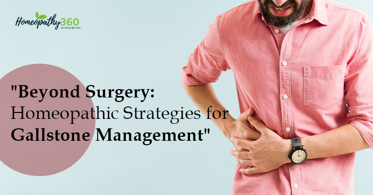
Abstract: Gallstone disease is a prevalent global health issue, with a rising incidence in recent years. The development of gallstones is influenced by a combination of genetic predisposition and environmental factors. While a significant portion of individuals with cholelithiasis remain asymptomatic, in symptomatic cases often resort to cholecystectomy, considered the gold standard treatment. However, these conventional treatments have inherent limitations and are associated with notable side effects. This article aims to explore homeopathic medicine as an alternative approach in the management of gallstones.
Keywords: Gallstones, Cholelithiasis, Homeopathic medicine and Sarcodes.
Introduction: Gallstones are hardened pebble-like particles, accumulate within the gallbladder and are primarily formed in gallbladder, although less frequently in intrahepatic or extrahepatic bile ducts. More patients with cholelithiasis have no obvious symptoms. Symptomatic patients presented with dyspepsia and biliary colic caused mainly by obstruction of the cystic duct.
Gallstones can give rise to severe complications, including cholecystitis, acute cholangitis, and pancreatitis.
Epidemiology: As of a comprehensive 2023 study featured in the journal Nature, the global prevalence of gallstones is projected to range between 10 – 15%. This implies that approximately 10-15% of the world’s population is affected by gallstones. Notably, the incidence of gallstones is markedly higher in women, with an estimated prevalence of 15-20%, in contrast to 5-10% in men.
Interestingly, gallstones are less frequently observed in regions such as India, the Far East, and Africa.
Gallstones are common with prevalences as high as 60% to 70% in American Indians and 10% to 15% in white adults of developed countries.
Ethnic differences abound with a reduced frequency in black Americans and those from East Asia, while being rare in sub-Saharan Africa.
Riskfactor: ‘ 4f ’ – female
forty fatty fertile
- Genes: A Swedish twin study showed that an inherited predisposition is responsible for 25% of the overall risk of developing gallstones . Lithogenic genes 1 and 2 (lith1 and lith2), playing a role in liver cholesterol secretion and regulating bile flow, have been described in murine models.
- Age : Risk of gall stones increases with advancing age. The incidence increase above the age of 40 yrs.
- Gender: cholelithiasis is more common in female than male. The impact of the higher incidence of cholelithiasis among related to the production of hormones, especially estrogen. Estrogens bound to estrogen receptors (ers) in the liver and increase the secretion of cholesterol intothe bile, promoting the formation of gallstones.
- Oral contraceptive : use by women may predispose to a more frequent occurrence of cholelithiasis.
- Obesity: obesity has been found to increase the risk of cholelithiasis development due toimpaired gallbladder motility, excessive hepatic secretion, and bile saturation of cholesterol
- Diet –the increased fat intake with highly refined sugars, fructose, and low fiber contents predisposes to the development of gallstones.
- Gastro intestinal disease: such as crohn’s disease, primary biliary cirrhosis, ilial resection etc are associated with interruption in enterohepatic circulation lead to gall stone formation.
- Clofibrate therapy: This Drug Is Used To Excrete Cholesterol In Hyper Lipoprotenemial There For Increases Concentration Of Cholesterol In Bile.
Pathogenesis of gallstone formation:
Cholesterolemia (excess cholesterol in blood)
Hyperbilirubinemia Gallbladder stasis
(excess bilirubin in blood) ( Impaired/decease gallbladder motility)
Classification: Based on their composition, gallstones are classified into :
- Pure Pigment stones are mainly observed in hemolytic diseases.Multiple, small, jet black, mulberry-shaped.
- Pure Cholesterol stones: Solitary, oval , large , smooth , yellow white.
- Pure Calcium carbonate: multiple, small, grey, grey white, hard.
- Mixed Gall Stone: Cholesterol bile pigment, calcium salt in varying combination. Multiple, variable size, Multifaceted.
Clinical features:-
- Asymptomatic : More than 80% of patients with gallstones are asymptomatic, characterizing gallstone disease as a predominantly silent disease.
- Symptomatic : In symptomatic patients, the manifestation of symptoms is attributed to stone-triggered inflammation and obstruction of the cystic and common bile ducts.
- Billiay colic – sudden and severe pain occur 1-2 hours after meal last for several hours.
- Pain in right upper quadrent (RUQ) and epigastrium, radiate to interscapular region or the tip of right scapula and back.
- Gall stone dyspepsia – pain with fatty food intolerance and flatulence.
- Pain associated with nausea and vomiting.
- Charcot’s triad – Associated with common bile duct obstruction and cholangitis.
Pain in Right upper quadrant
SIGN:-
Jaundice Fever
- MURPHY’S SIGN: Firmly palpate the RUQ region and ask the patient to take deep breath. A positive sign is when significant pain is elicited by this maneuver, usualy stopping them mid breath.
- BOA’S SIGN – An Area No Hyper Aesthesia Between 9th & 11th Rib Posteriorly Right Side.
COMPLICATIONS-
- Cholecystitis
- Cholangitis
- Choledocholothiasis
- Porcelain gall bladder
- Gall blader polyp
- Gall bladder fistulae
- Acute pancreatitis
- Gallstone ileus
- Mirrizzi’s syndrome (inflammation of common bile duct by a gall stone in the cystic duct.)
- Cholangiocarcinoma (cholelithiasis is not clearly a predisposing factor for cholangiocarcinoma. – harrison.
- Diagnosis based on history and physical examination with confirmatory radiological studies.
INVESTIGATION :
- ray Ultrasound CT scan
ERCP (Endoscopic retrograde cholangiopancreatography) Cholescintigraphy
HOMOEOPATHIC MANAGEMENT
- BERBERIS VULGARIS: – suited to bilious diathesis. Act on liver promote flow of bile. Stitches in the region of gall-bladder; worse, pressure, extending to stomach. Catarrh of the gall-bladder with constipation and yellow complexion. Bubbling sensation, or stitches marked. Sticking pain in the region of liver and gall-bladder shooting to left shoulder, < by pressure. Gall-stone colic, followed by jaundice. Aching and
shooting pain in epigastrium and hepatic region. Colic, billiary arresting breathing.
- CALCAREA CARBONICUM:- Gall-stone colic. Increase of fat in abdomen. Sensitive to slightest pressure. Cannot bear tight clothing around abdomen. Liver region painful agg. Stooping. Patient can be describe as fat, flabby, fair, forty, perspiring, cold and damp.
- CARDUUS MARINUS: – Act on liver and portal system, Causing soreness, pain, jaundice. Pain in region of liver. Left lobe very sensitive. Fullness and soreness. Swelling of gall bladder with painful tenderness. Gall stone disease with enlarged liver in transverse direction (chelidonium- more vertical).
- CHELIDONIUM MAJUS: – Predominantly right sided remedy. Gallstones-colic, pain through stomach to back and right shoulder-blade. The jaundiced skin and constant pain under inferior angle of right scapula. Tongue yellow, with imprint of teeth; large and flabby. Taste bitter, want hot food and drink. Eating gives temporarily relieves. Bilious complication.
- CHINA OFFICINALIS:- Gall-stone colic. Shooting and pressive pain in right hypochondria and hepatic region, esp. when touched. Much flatulent colic; better bending double. Periodicity is most marked. Tympanitic abdomen. Jaundice.
- CHIONANTHUS VIRGINICA: – Gall stones. Hepatic region tender. Sore, enlarged, with jaundice and constipation. Hot, Bitter eructation, tasting as if food were fermenting in stomach. Vomiting of very dark green, ropy, and exceedingly bitter bile. clay- coloured, soft, yellow, pasty stool. Tongue heavily coated. No appetite. Billious colic. Cholecystitis.
- DIOSCOREA VILLOSA: – Gall stone colic. Pain from gall-bladder to chest, back, and arms; worse, bending forewords and while lying; better on standing erect, or bending backwards and by walking about. Pains from liver, shooting upward to right nipple. Pains Are Unbearable, Sharp; Cutting, Twisting, Griping, Grinding, that radiate to distant parts; they occur in paroxysm. Violent twisting colic, occurring in paroxysm, as if intestine were grasped and twisted by a powerful hand. Rumbling, with emission of much flatus.
- LYCOPODIUM: – Predominantly right sided remedy. Suited to ailments gradually developed with failure of digestive power. Violent Gall stone colic-sharp pain in dorsal hepatic region, in Rt. shoulder and arm. Fermentation in abdomen, which produce a loud croaking sound. Gurgling and borborygmi in abdomen, esp. on lower abdomen not relieved by belching. Sour vomiting and eructation. Canine hunger but a few mouthfuls fill up to the throat. Worse in afternoon, at night, from 4 to 8pm.
- NUX VOMICA: – Weight worse after eating nausea & vomiting with much retching. Nausea; amel. If he can vomit. Region of stomach very sensitive to pressure. Colic with upward pressure, causing short breath.
Bruised soreness of abdominal walls. Gallstones colic. Jaundice; from anger aggravation.
SARCODES USED FOR GALL STONES MANAGEMENT: –
- CHOLESTERINUM – Gall stone, jaundice and insomnia obstinate hepatic ingoargments. Cholestrenum is the physiological opponent of lecithin.
- FEL TAURI (OX-GALL,TRITURATION OF GALL BLADDER)– Liquefies bile and act as a purgatives and chologue obstruction of gall duct. Billiary calculi. Jaundice. Emulsify fats, increases the peristaltic action of intestine & duodenal secretion. Eructation, gurgling in stomach and epigastric region. Tendency to sleep after eating.
DOSE:- Lower Trituration
REFERENCES:-
| 1. | Davidson S. Davidson’s Principles and Practice of Medicine. 23rd ed. Stuart H Ralston IDPMWSRPH, editor. New York: Elsevier Ltd; 2018. |
| 2. | Harsh Mohan. Textbook of Pathology. 7th ed. Jaypee Brothers medical publisher (P) Ltd; New Delhi. 2015 |
| 3. | Stinton, L. M., Myers, R. P., & Shaffer, E. A. (2010). Epidemiology of gallstones. Gastroenterology clinics of North America, 39(2), 157 –vii. |
| 4. | Boericke W. Boericke’s New Manual of Homoeopathic Materia Medica withREPERTORY. third revised edition and Augmented edition based on Ninth edition ed. USA: B. Jain Publisher (P) Ltd; 2016. |
| 5. | PHATAK SR. Materia medica of Homoeopathic Medicine Noida: B.Jain Publishers (P)LTD; 2019. |
| 6. | Clarke, J.H. (1904) A dictionary of practical Materia Medica : A clinical repertory to the dictionary. London. |
| 7. | Patil, J.D. Group study in Homoeopathic Materia Medica B.Jain Publishers(p) LTD; New Delhi 2018 |


