Eyeballs are cystic, almost spherical structures resembling fluid filled balls. They are kept distended by the pressure of the fluid inside them.
DIMENSIONS
The anterior part of these spheres consists of the cornea which has a radius of about 7.8 mm whereas the posterior part consists of the sclera which has a radius of about 12 mm. The junction between the cornea and sclera is called Limbus and the conjunctiva is very firmly attached here.
Other measurements include-
Anteroposterior diameter- 24mm
Horizontal Diameter- 23.5mm
Vertical Diameter- 23mm
Circumference- 75mm
Volume- 6.5 ml
Weight- 7gm
Since the eyeballs are spherical structures, the central point on the maximal convexities of the anterior and posterior curvatures of the eyeball are called anterior and posterior poles respectively and the midplane between the two poles is known as the equator.
You may read more on the anatomy of the eyeball in B D Chaurasia’s Human Anatomy Volume 3 which features With Newly drawn diagrams, redesigned on the basis of reader–teacher perceptions, in vivid colours, imposing dimensions, with clarity of depiction, using fresh labelling, this book is essential for all students looking for an enhanced and well illustrated clinical anatomy for effective learning during the clinical years.
STRUCTURES LINING THE EYEBALL
There are three layers that bound the eyeball from within. These are-
Inner Nervous Coat – It is concerned with visual functions and projects to the visual cortex through the visual pathway.
Middle Vascular Coat – It supplies nutrition to the various structures of the eyeball and consists of iris, ciliary body and choroid from anterior to posterior.
Outer Fibrous Coat- A dense and strong wall which protects the intraocular structures and consists of the transparent Cornea on the anterior side and opaque sclera which forms the 5/6th posterior part.
CHAMBERS OF THE EYEBALL
The eyeball is divided into two segments –
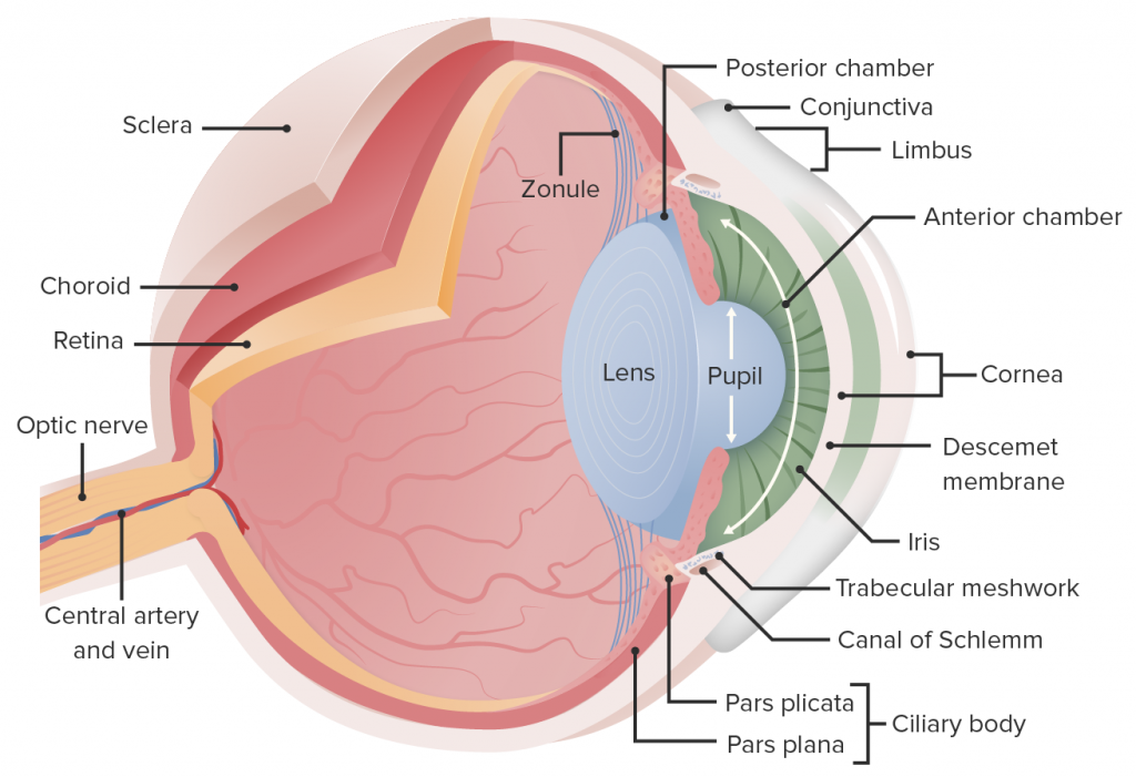
- Anterior Segment- It includes the crystalline lens (suspended from the ciliary body by zonules) and structures anterior to it namely iris, cornea and two aqueous humour filled spaces called anterior and posterior chambers.
- Anterior chamber : It is bounded anteriorly by the back of the cornea and posteriorly by the anterior surface of iris and part of the ciliary body. This chamber is about 2.5 mm deep in the centre for healthy adults and contains about 0.25 ml of aqueous humour.
- Posterior Chamber : A triangular space containing 0.06 ml of aqueous humour, this is bounded anteriorly by the posterior surface of the iris and part of ciliary body, posteriorly by the crystalline lens and its zonules and laterally by the ciliary body.