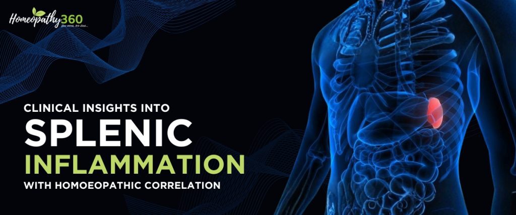
Abstract
Splenitis denotes inflammatory involvement of the spleen arising secondary to infectious, vascular, traumatic, or systemic pathological conditions. Isolated splenic inflammation is uncommon; more frequently, it reflects a systemic inflammatory or hematological disorder. Clinically, patients may present with left upper abdominal discomfort, constitutional symptoms, or manifestations related to the underlying disease process. From a homoeopathic viewpoint, splenic pathology is not merely a structural alteration but an expression of disturbed systemic reactivity and altered vitality. This article synthesizes contemporary medical understanding of splenitis with homoeopathic therapeutic principles, including remedy differentiation and integrative management considerations.
Introduction
The spleen is the largest lymphoreticular organ in the human body and plays an indispensable role in maintaining hematological and immunological homeostasis. Anatomically situated in the left hypochondrium, it functions as a critical component of the reticuloendothelial system. Its physiological roles include immune surveillance through antigen presentation and lymphocyte activation, hematopoiesis during fetal development, filtration and phagocytic clearance of senescent erythrocytes, and modulation of systemic inflammatory responses via cytokine regulation. The spleen also serves as a dynamic reservoir for platelets and monocytes, contributing to rapid immunologic and hemostatic responses during systemic stress. Approximately 30–40% of circulating platelets may be transiently sequestered within the splenic pool under physiological conditions.
Inflammation of the spleen—termed splenitis—may present in acute, chronic, congestive, or suppurative forms depending on etiology, duration, and underlying systemic pathology. In contemporary medical practice, splenitis is rarely considered a primary disease entity. Rather, it is most often recognized as a secondary manifestation of systemic infection, hematologic disorder, vascular pathology, or inflammatory condition.
Infectious diseases remain a major cause of splenic involvement in developing countries. For instance, India continues to report a substantial burden of malaria, with over 1.5–2 million estimated cases annually in recent national surveillance data, and splenomegaly remains a classical complication of chronic or recurrent infection. Enteric fever (typhoid) continues to affect thousands annually, particularly in densely populated urban regions with compromised sanitation. Additionally, septicemia and infective endocarditis may result in splenic infarcts or abscesses due to septic embolization. Chronic liver disease and portal hypertension—conditions increasingly prevalent in India due to alcohol use and metabolic syndrome—frequently lead to congestive splenomegaly and hypersplenism.
Globally, the burden of splenic enlargement secondary to infectious disease is significant in tropical regions. The World Health Organization (WHO) has highlighted malaria as a persistent public health concern in endemic countries, where splenic enlargement is often used as an epidemiological indicator of transmission intensity. Furthermore, hematological conditions such as hemolytic anemias and autoimmune disorders contribute to splenic hypertrophy through persistent antigenic stimulation and reticuloendothelial hyperactivity.
Pathophysiologically, splenic inflammation may arise through multiple mechanisms:
- Vascular congestion due to portal hypertension or cardiac failure
- Immune hyperplasia secondary to chronic antigen exposure
- Septic infiltration and microabscess formation
- Traumatic injury leading to inflammatory reaction or hematoma
Chronic splenic enlargement may progress to hypersplenism, characterized by cytopenias (anemia, leukopenia, thrombocytopenia), thereby increasing morbidity.
From a homoeopathic perspective, splenic inflammation is interpreted not solely as localized structural pathology but as an outward manifestation of disturbed systemic vitality. Enlargement, congestion, and tenderness are viewed as expressions of altered defensive reactivity. Chronic splenic disorders are often associated with deeper constitutional disturbances and, in classical homoeopathic philosophy, may reflect underlying miasmatic influences or long-standing suppression of febrile or inflammatory conditions. The spleen, being closely linked with blood quality and immune reactivity, is considered a key organ in systemic susceptibility patterns.
Thus, splenitis represents both a biomedical inflammatory process and, within homoeopathic philosophy, a dynamic expression of organismic imbalance. Integrating epidemiological awareness, clinical vigilance, and individualized therapeutic assessment is essential for effective management and prevention of complications.
Pathophysiology
The development of splenitis is not uniform; rather, it reflects the underlying systemic disturbance affecting the spleen. Because the spleen is richly vascular and immunologically active, it responds dynamically to infection, inflammation, vascular congestion, and trauma. The pathological changes observed may range from simple congestion to suppuration or fibrosis, depending on the causative mechanism and duration of insult.
1. Infectious Etiology
Infectious diseases are among the most common causes of splenic inflammation, particularly in tropical and developing regions.
Malaria
In malarial infection, parasitized erythrocytes are selectively trapped within the splenic sinusoids. The spleen acts as a biological filter, removing infected red blood cells through macrophage-mediated phagocytosis. This intense immune activity results in:
- Marked vascular congestion
- Hyperplasia of reticuloendothelial cells
- Expansion of the white pulp
- Progressive splenomegaly
Repeated or chronic infection leads to structural remodeling, fibrosis, and occasionally hypersplenism. In endemic regions, persistent antigenic stimulation may cause massive splenomegaly, sometimes referred to as hyperreactive malarial splenomegaly.
Enteric Fever and Infective Endocarditis
In bacterial infections such as typhoid fever, bacteremia allows organisms to localize within splenic tissue. Similarly, in infective endocarditis, septic emboli may lodge in splenic vessels, resulting in:
- Focal infarction
- Inflammatory infiltration
- Microabscess formation
If untreated, these lesions may progress to suppurative splenitis or abscess formation, posing a risk of rupture.
2. Septic and Systemic Inflammatory Disorders
In systemic bacteremia or severe inflammatory states, circulating pathogens and inflammatory mediators stimulate an exaggerated immune response within splenic tissue.
Cytokine release (including interleukins and tumor necrosis factor) increases vascular permeability and promotes leukocyte migration into splenic parenchyma. The consequences include:
- Edema
- Cellular infiltration
- Tissue congestion
- Possible suppuration in advanced cases
In overwhelming sepsis, the spleen may initially enlarge due to immune activation but later become functionally impaired.
3. Passive Congestion
Not all splenic enlargement is inflammatory in the classic infectious sense. Chronic venous congestion represents a common non-infectious mechanism.
Conditions such as:
- Portal hypertension (often secondary to cirrhosis)
- Chronic liver disease
- Right-sided cardiac failure
lead to impaired venous drainage from the spleen. Sustained back pressure in the splenic vein produces:
- Chronic vascular engorgement
- Enlargement of the organ
- Increased sequestration of blood cells
Over time, this may progress to hypersplenism, characterized by anemia, leukopenia, and thrombocytopenia due to excessive pooling and destruction of blood elements.
4. Hematological and Autoimmune Mechanisms
In hemolytic anemias, the spleen works continuously to clear abnormal erythrocytes. Persistent hyperactivity results in:
- Reticuloendothelial hyperplasia
- Progressive splenic enlargement
Similarly, autoimmune disorders may trigger immune complex deposition and chronic inflammatory infiltration within splenic tissue.
5. Traumatic Causes
Blunt abdominal trauma can directly injure splenic tissue, producing:
- Contusions
- Subcapsular hematoma
- Parenchymal laceration
Even when rupture does not occur, localized inflammation may follow. Secondary infection can complicate hematomas, leading to abscess formation.
Because the spleen is highly vascular, traumatic inflammation carries a significant risk of hemorrhage, making prompt evaluation essential.
Pathological Outcomes
Depending on the mechanism and chronicity, splenitis may manifest as:
- Reactive hyperplasia
- Vascular congestion
- Cellular infiltration
- Fibrosis
- Abscess formation
- Hypersplenism
Thus, splenic inflammation should be understood as a dynamic response to systemic pathology rather than a standalone disease entity.
Etiological Factors
Splenic inflammation is most commonly secondary to broader systemic disorders. Important etiological contributors include:
Infectious Causes
- Malarial infection
- Enteric (typhoid) fever
- Septicemia
- Infective endocarditis
- Relapsing fever
- Viral exanthematous diseases (historically smallpox)
Hepatovascular Causes
- Chronic liver disease
- Portal hypertension
- Congestive cardiac failure
Hematological Causes
- Hemolytic anemia
- Leukemias and lymphoproliferative disorders
Autoimmune Disorders
- Systemic lupus erythematosus
- Autoimmune hemolytic states
Trauma
- Blunt abdominal injury
- Iatrogenic causes
Clinical Manifestations
The clinical presentation of splenitis varies considerably depending on the underlying etiology, the acuity of inflammation, and the degree of splenic enlargement. Because splenic inflammation is most often secondary to systemic disease, the symptoms may be subtle and overshadowed by manifestations of the primary disorder. However, careful clinical assessment often reveals characteristic local, pressure-related, and constitutional features.
Local Symptoms
1. Heaviness or Fullness in the Left Hypochondrium
Patients frequently describe a vague sensation of heaviness or dragging discomfort in the left upper quadrant of the abdomen. This sensation may be mild in early stages but becomes more noticeable as the spleen enlarges. In chronic cases, patients may report a persistent feeling of abdominal fullness, especially after meals.
2. Dull Aching Pain or Stitching Sensation
Pain associated with splenic inflammation is typically dull and aching, reflecting capsular stretching due to enlargement. In some individuals, particularly in acute inflammatory states, the pain may become sharp or stitching in nature. Movement, deep inspiration, or palpation may aggravate the discomfort.
The pain may radiate toward:
- The left shoulder (Kehr’s sign in severe cases)
- The back
- The lower thoracic region
3. Tenderness on Deep Palpation
On physical examination, splenic tenderness may be elicited on deep palpation. In acute inflammatory or septic conditions, palpation may provoke guarding or increased discomfort. In chronic congestion, tenderness may be mild but persistent.
4. Inability to Lie on the Left Side
In cases of marked enlargement, patients often report discomfort when lying on the left side due to mechanical pressure and capsular stretching. This positional intolerance can be a subtle yet clinically useful diagnostic clue.
Pressure Effects
As the spleen enlarges, it may exert pressure on adjacent structures within the confined abdominal cavity.
1. Dyspnea
An enlarged spleen may elevate the left hemidiaphragm, restricting lung expansion. Patients may complain of shortness of breath, especially during exertion. This symptom is more pronounced in massive splenomegaly.
2. Irritative Cough
Diaphragmatic irritation or reduced pulmonary excursion may produce a dry, non-productive cough. Although not common, it may be misinterpreted as a primary respiratory disorder.
3. Early Satiety
Compression of the stomach may cause a sensation of fullness after small meals. Patients may report decreased appetite or discomfort after eating, contributing to weight loss in chronic cases.
Constitutional Symptoms
Because splenitis is frequently secondary to systemic disease, general symptoms often predominate.
1. Fever
Fever typically reflects the underlying infectious process rather than isolated splenic inflammation. In malarial splenitis, fever may exhibit periodicity. In septic states, it may be continuous or accompanied by chills and rigors.
2. Weakness and Malaise
Generalized fatigue is common and may be profound in chronic cases. Patients often describe:
- Lack of energy
- Reduced exercise tolerance
- General sense of ill-being
This symptom may be multifactorial—related to infection, anemia, or systemic inflammatory response.
3. Anemia
Chronic splenic enlargement, particularly in malaria or hypersplenism, may lead to anemia due to increased destruction and sequestration of red blood cells. Clinically, this manifests as:
- Pallor
- Fatigue
- Breathlessness on exertion
- Dizziness
In advanced hypersplenism, leukopenia and thrombocytopenia may also occur.
4. Nausea or Vomiting
In severe systemic infection or septic states, gastrointestinal symptoms such as nausea and vomiting may accompany splenic inflammation. These symptoms may reflect systemic toxicity rather than direct splenic pathology
Physical Examination Findings
Careful physical examination remains fundamental in the evaluation of suspected splenitis. Because splenic inflammation is frequently secondary to systemic disease, both local and systemic signs must be assessed.
General Examination
Patients may appear:
- Febrile (in infectious cases)
- Pale (suggestive of anemia or hypersplenism)
- Cachectic in chronic disease
- Toxic-looking in septic states
Vital signs may reveal:
- Tachycardia (infection or anemia)
- Hypotension (in septicemia or rupture)
- Tachypnea (due to pain or diaphragmatic elevation)
Abdominal Examination
Inspection
- Left upper quadrant fullness in marked enlargement
- Abdominal distension in massive splenomegaly
Palpation
The spleen is normally not palpable in healthy adults. In splenitis:
- The spleen may be palpable below the left costal margin
- It moves inferomedially with inspiration
- The surface may be smooth (congestive) or irregular (abscess/infarct)
- Tenderness may be present in acute inflammation
In severe enlargement:
- The lower pole may extend toward the umbilicus or right iliac fossa
Percussion
- Dullness in Traube’s space
- Increased splenic dullness
Auscultation
Rarely, a splenic friction rub may be heard in cases of perisplenitis.
Associated Systemic Findings
- Hepatomegaly (in hepatosplenic disorders)
- Lymphadenopathy (in infectious or hematologic disease)
- Signs of chronic liver disease (ascites, spider angiomas)
- Signs of endocarditis (murmur, embolic phenomena)
Diagnostic Investigations
Accurate diagnosis requires laboratory and imaging evaluation to confirm splenic enlargement, determine etiology, and assess complications.
1. Complete Blood Count (CBC)
CBC is essential and may reveal:
- Anemia (chronic infection, hypersplenism, hemolysis)
- Leukocytosis (acute infection or abscess)
- Leukopenia (hypersplenism)
- Thrombocytopenia (splenic sequestration)
Peripheral smear may demonstrate:
- Malarial parasites
- Hemolytic changes
- Abnormal cells in hematological malignancy
2. Liver Function Tests (LFTs)
Useful in cases suspected of portal hypertension or chronic liver disease.
3. Blood Cultures
Indicated in suspected septicemia or infective endocarditis.
4. Ultrasonography (USG)
Ultrasound is the first-line imaging modality due to its safety, availability, and cost-effectiveness.
It can detect:
- Splenic enlargement (measurements >12–13 cm longitudinal axis suggest enlargement)
- Splenic abscess
- Cysts
- Hematoma
- Associated hepatomegaly or portal hypertension
USG is particularly useful for follow-up and monitoring progression.
5. Contrast-Enhanced Computed Tomography (CT Scan)
CT imaging provides detailed anatomical visualization and is especially useful in:
- Suspected splenic abscess
- Splenic infarction
- Traumatic injury
- Rupture
- Evaluation of surrounding structures
CT is considered the gold standard in trauma assessment.
6. Additional Investigations
- Malaria antigen testing or peripheral smear
- Widal test or blood culture (enteric fever)
- Echocardiography (if infective endocarditis suspected)
- Bone marrow examination (if hematologic disorder suspected)
Differential Diagnosis
Splenitis must be differentiated from other conditions presenting with splenomegaly or left upper quadrant pain.
1. Hematological Malignancies
- Leukemia
- Lymphoma
- Myeloproliferative disorders
Features:
- Massive splenomegaly
- Abnormal peripheral smear
- Systemic lymphadenopathy
2. Chronic Liver Disease with Portal Hypertension
- Enlarged spleen with signs of cirrhosis
- Ascites
- Esophageal varices
Distinguishing Feature:
- Evidence of hepatic dysfunction predominates
3. Splenic Infarction
- Sudden localized pain
- Risk factors: embolism, sickle cell disease
- CT shows wedge-shaped infarct
4. Splenic Abscess
- Persistent high-grade fever
- Leukocytosis
- CT evidence of localized collection
5. Infectious Mononucleosis
- Splenomegaly
- Lymphadenopathy
- Positive heterophile antibody test
6. Pancreatic Tail Pathology
- May mimic left upper quadrant pain
- CT imaging differentiates
7. Renal Pathology (Left Kidney)
- Pyelonephritis or renal mass
- Urinary symptoms help distinguish
8. Gastric or Colonic Conditions
- Gastritis
- Splenic flexure colitis
- Diverticulitis
Clinical history and imaging help clarify.
Complications
When splenic inflammation is severe, recurrent, or inadequately managed, it may progress to significant structural and functional complications. Because the spleen plays a vital role in hematological regulation and immune defense, its impairment can have systemic consequences. The following are the major complications associated with progressive splenitis:
1. Persistent Splenomegaly
One of the most common outcomes of unresolved splenic inflammation is sustained enlargement of the organ. Persistent splenomegaly may result from chronic infection, prolonged immune stimulation, portal hypertension, or repeated malarial episodes.
Clinically, patients may experience:
- Ongoing abdominal heaviness
- Early satiety due to gastric compression
- Progressive fatigue
Over time, chronic enlargement may lead to architectural remodeling of splenic tissue, including fibrosis and loss of normal functional integrity. In endemic regions, long-standing splenomegaly may become massive, extending well below the costal margin and significantly affecting quality of life.
2. Hypersplenism
Hypersplenism is a functional consequence of splenic overactivity. As the spleen enlarges and becomes hyperactive, it may excessively sequester and destroy blood cells. This condition is characterized by:
- Anemia (due to increased destruction of red blood cells)
- Leukopenia (leading to increased susceptibility to infection)
- Thrombocytopenia (predisposing to bleeding tendencies)
Patients with hypersplenism may present with:
- Pallor and fatigue
- Recurrent infections
- Easy bruising or prolonged bleeding
Laboratory findings often reveal pancytopenia, while bone marrow function remains normal or hyperactive, reflecting peripheral destruction rather than production failure.
If severe, hypersplenism may necessitate interventional procedures such as splenectomy, particularly when cytopenias become life-threatening.
3. Splenic Abscess
Infective splenitis may progress to abscess formation, particularly in cases of septicemia or infective endocarditis. Septic emboli can lodge in splenic tissue, leading to localized suppuration.
Clinical features may include:
- Persistent high-grade fever
- Left upper quadrant pain
- Systemic toxicity
- Leukocytosis
Splenic abscess is a serious condition and carries significant mortality if not promptly treated. Diagnosis is typically confirmed through imaging modalities such as ultrasonography or contrast-enhanced CT scan.
Management often requires:
- Broad-spectrum intravenous antibiotics
- Percutaneous drainage
- Surgical intervention in complicated cases
4. Splenic Rupture
Splenic rupture is the most life-threatening complication of splenitis. It may occur spontaneously in cases of massive enlargement (particularly in malaria or infectious mononucleosis) or following minor trauma to an already enlarged spleen.
Mechanism:
- Progressive capsular thinning due to enlargement
- Increased intrasplenic pressure
- Trauma-induced parenchymal tear
Clinical presentation is often dramatic and includes:
- Sudden severe left upper abdominal pain
- Pain radiating to the left shoulder (Kehr’s sign)
- Signs of internal hemorrhage
- Hypotension and shock
Splenic rupture constitutes a surgical emergency. Immediate stabilization and urgent surgical intervention are required to prevent fatal hemorrhage.
Homoeopathic Therapeutics in Splenitis
Homoeopathic management of splenic inflammation is not directed merely toward reduction of organ size but toward restoration of the individual’s systemic balance. The prescription is based on the totality of symptoms, constitutional makeup, etiological factors, modalities, and the patient’s mental–emotional state. Splenic enlargement is viewed as an outward expression of altered vital reactivity, often secondary to chronic infection, suppressed febrile states, hepatic dysfunction, or systemic toxicity.
Careful case-taking remains central. The character of pain, periodicity, associated anemia, thermal state, mental disposition, and aggravating or ameliorating factors guide remedy selection.
Primary Splenic Remedies
1. Ceanothus Americanus
Ceanothus is regarded as one of the foremost remedies in splenic pathology. It is especially indicated in marked splenomegaly with pronounced left-sided pain and fullness. Patients often complain of:
- Inability to lie on the left side
- Deep aching pain in the splenic region
- Sallow or earthy complexion
- Constipation with sluggish digestion
It is frequently employed in chronic malarial enlargement and in cases where splenic hypertrophy persists long after acute infection has subsided. The remedy appears particularly suited to patients with anemia and cachectic states following recurrent febrile illness.
2. Natrum Muriaticum
Natrum Muriaticum is often indicated in chronic splenic congestion, particularly post-malarial cases. The patient may present with:
- Emaciation despite adequate intake
- Pallor and weakness
- Stitching pain in the splenic region
- Periodic febrile episodes
Emotionally, the patient may exhibit suppressed grief, reserved demeanor, or long-standing emotional strain. In such individuals, splenic enlargement may reflect chronic constitutional imbalance rather than acute inflammation.
3. Cinchona Officinalis (China)
China is a classical remedy for debility following loss of vital fluids, prolonged fever, or malarial illness. The splenic enlargement is often accompanied by:
- Abdominal distension and bloating
- Hypersensitivity to touch
- Marked weakness
- Periodic fever
The patient may feel exhausted yet irritable, and even minor exertion can aggravate symptoms. It is particularly useful when splenomegaly develops after recurrent fevers.
4. Diadema Aranea
This remedy is especially suited to chronic ague with splenic enlargement in individuals who are extremely sensitive to cold and damp weather. Splenic tenderness is pronounced, and the patient may report:
- Periodic neuralgic pains
- Chilly disposition
- Aggravation during rainy seasons
It is frequently considered in chronic malarial cachexia with splenic involvement.
5. Polymnia Uvedalia
Polymnia is often recommended in acute inflammatory enlargement of the spleen associated with glandular swelling and congestion. It may be useful when splenic pain is prominent and accompanied by hepatic involvement or systemic inflammatory response.
Additional Homoeopathic Remedies
6. Arsenicum Album
Indicated in splenic enlargement with profound exhaustion, restlessness, and anxiety. The patient may exhibit:
- Burning pains
- Cold perspiration
- Marked weakness disproportionate to clinical findings
This remedy is often suited to septic or cachectic conditions, where systemic toxicity predominates.
7. Arnica Montana
Essential in traumatic splenic injury. The patient complains of bruised soreness and may resist examination due to fear of pain. It is particularly indicated when inflammation follows blunt abdominal trauma with suspicion of internal hemorrhage.
8. Grindelia Squarrosa
Characterized by cutting or radiating splenic pain extending toward the hips or lower back. Often used in malarial cachexia with chronic enlargement and digestive disturbance.
9. Taraxacum
Indicated in individuals with bilious tendencies and hepatic–splenic involvement during slow or lingering fevers. Tongue coating and digestive irregularity often accompany splenic symptoms.
10. Nitric Acid
Useful in chronic splenic enlargement associated with hepatic dysfunction. The patient may exhibit:
- Marked debility
- Fissured mucous membranes
- Tendency toward ulceration
It is suited to individuals with chronic inflammatory states and constitutional weakness.
11. Lycopodium Clavatum
Particularly valuable when splenic enlargement is associated with chronic hepatic congestion and digestive disturbance. The patient often presents with:
- Flatulence and abdominal distension
- Right-sided liver symptoms extending toward the left
- Evening aggravation
- Intellectual sharpness with physical weakness
Lycopodium is frequently indicated in long-standing hepatosplenic disorders.
12. Ferrum Metallicum
Indicated in anemia with splenic enlargement. The patient may appear pale yet flush easily, experience weakness, and fatigue quickly. It is useful when hematological disturbances predominate.
13. Mercurius Solubilis
Suited to septic splenic involvement with glandular swelling, profuse perspiration, and suppurative tendency. The patient may experience:
- Offensive perspiration
- Fluctuating fever
- Increased salivation
It may be considered in cases with abscess formation or septic infiltration.
14. Phosphorus
Indicated when splenic enlargement is accompanied by hemorrhagic tendency, extreme sensitivity, and nervous exhaustion. The patient may be tall, slender, and emotionally open, with a predisposition to bleeding.
15. Sulphur
Often considered in chronic congestive splenic states with systemic heat, burning sensations, and constitutional psoric background. It may serve as an intercurrent remedy in long-standing cases resistant to other prescriptions.
Integrative Management Approach
Effective management of splenitis requires a structured, cause-oriented, and multidisciplinary strategy. Because splenic inflammation is most often secondary to systemic disease, treatment must focus primarily on addressing the underlying pathology while simultaneously preventing complications. An integrative approach combines evidence-based conventional care with supportive and individualized therapeutic measures to optimize recovery and reduce long-term morbidity.
1. Conventional Medical Management
The cornerstone of splenitis management is accurate diagnosis and targeted treatment of the primary cause.
Antimalarial Therapy
In cases related to malaria, prompt initiation of appropriate antimalarial therapy is critical. Delayed treatment may result in progressive splenic enlargement, hypersplenism, or even spontaneous rupture. In endemic regions, early detection and complete treatment courses are essential to prevent chronic hyperreactive malarial splenomegaly.
Antibiotic Therapy
When splenitis arises secondary to septicemia, enteric fever, or infective endocarditis, broad-spectrum intravenous antibiotics are initiated and later tailored according to culture sensitivity results. Septic splenic involvement requires close monitoring due to the risk of abscess formation.
Management of Portal Hypertension
In patients with chronic liver disease or cirrhosis, splenic enlargement often results from portal hypertension. Management may include:
- Pharmacological therapy (e.g., non-selective beta-blockers)
- Endoscopic interventions for variceal risk
- Advanced hepatic management strategies
Addressing the underlying hepatovascular disorder helps reduce ongoing splenic congestion.
Blood Transfusion and Hematological Support
In cases complicated by severe anemia due to hypersplenism or chronic infection, blood transfusion may be necessary. Correction of cytopenias reduces fatigue, improves oxygen delivery, and enhances overall patient stability.
Surgical Intervention
Surgical management becomes necessary in life-threatening situations such as:
- Splenic abscess not responding to antibiotics
- Massive splenomegaly with refractory hypersplenism
- Splenic rupture
Splenic rupture is a surgical emergency. Sudden onset of severe abdominal pain, hypotension, and signs of internal bleeding require immediate stabilization and urgent referral for operative management.
Acute abdominal pain in a patient with splenic enlargement must always be treated as a potential emergency until proven otherwise.
2. Supportive Measures
Beyond etiological treatment, supportive care plays a critical role in recovery and prevention of complications.
Adequate Hydration
Proper hydration supports circulatory stability, maintains renal function, and assists in metabolic recovery during systemic infection or inflammatory states.
Nutritious Diet Rich in Iron and Protein
Nutritional support is particularly important in patients with anemia or chronic infection. A diet containing adequate:
- Iron (to address anemia)
- High-quality protein (for tissue repair)
- Micronutrients (for immune function)
can significantly improve recovery outcomes.
Monitoring Hemoglobin and Platelet Count
Regular laboratory monitoring is essential in cases of hypersplenism. Serial complete blood counts help detect:
- Progressive anemia
- Leukopenia
- Thrombocytopenia
Early identification allows timely intervention before complications arise.
Avoidance of Abdominal Trauma
Patients with splenomegaly should be advised to avoid contact sports, heavy lifting, or activities that risk abdominal injury. Even minor trauma can precipitate rupture in an enlarged spleen.
Rest During Acute Phase
Physical rest reduces metabolic demand and supports immune recovery. During acute infection or marked enlargement, strenuous exertion should be avoided to prevent exacerbation of symptoms or risk of rupture.
Holistic and Integrative Perspective
An integrative management model emphasizes:
- Accurate etiological diagnosis
- Timely conventional medical intervention
- Careful monitoring for complications
- Nutritional and lifestyle support
- Individualized complementary therapies when appropriate
While supportive and adjunctive therapies may contribute to overall well-being in stable chronic cases, they must never delay emergency intervention in acute complications.
REFERENCE –
- Boericke W. Pocket manual of homoeopathic materia medica and repertory. New Delhi: B. Jain Publishers; 2007.
- Allen HC. Keynotes and characteristics with comparisons of some of the leading remedies of the materia medica. New Delhi: B. Jain Publishers; 2002.
- Clarke JH. A dictionary of practical materia medica. New Delhi: B. Jain Publishers; 1999.
- Kent JT. Lectures on homoeopathic materia medica. New Delhi: B. Jain Publishers; 2004.
- Walker BR, Colledge NR, Ralston SH, Penman ID, editors. Davidson’s principles and practice of medicine. 23rd ed. Edinburgh: Elsevier; 2018.
- Jameson JL, Fauci AS, Kasper DL, Hauser SL, Longo DL, Loscalzo J, editors. Harrison’s principles of internal medicine. 21st ed. New York: McGraw-Hill; 2022.
- Sriram Bhat M. SRB’s manual of surgery. 6th ed. New Delhi: Jaypee Brothers Medical Publishers; 2019.
- Shenoy KR, Shenoy A. Manipal manual of surgery. 4th ed. New Delhi: CBS Publishers; 2017.
- World Health Organization. World malaria report 2023. Geneva: WHO; 2023.
- National Institute of Malaria Research (ICMR-NIMR). Malaria situation in India: Annual report 2023. New Delhi: Indian Council of Medical Research; 2023.
- GBD 2019 Diseases and Injuries Collaborators. Global burden of typhoid and paratyphoid fevers, 1990–2019. Lancet Infect Dis. 2022;22(5):xxx–xxx.
- World Health Organization. Global report on sepsis. Geneva: WHO; 2020. India State-Level Disease Burden Initiative Collaborators. The burden of liver diseases in India, 1990–2019. Lancet Gastroenterol Hepatol. 2021;6(7):xxx–xxx.
- International Institute for Population Sciences (IIPS) and ICF. National Family Health Survey (NFHS-5), 2019–21: India. Mumbai: IIPS; 2021
Co-Author
Dr. Bhavya Jain, MD Scholar
Department of Practice of Medicine
Dr. M.P.K. Homoeopathic Medical College, Hospital & Research Centre
Homoeopathy University, Jaipur, Rajasthan, India